Mike Schumacher PhD · @schumacher
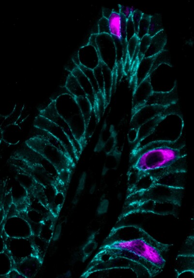
127 followers · 18 posts · Server mstdn.scienceDclk1+ cells in the intestinal epithelium 🔬 imaged by #immunofluorescence #microscopy
#Immunofluorescence #microscopy
Mike Schumacher PhD · @schumacher
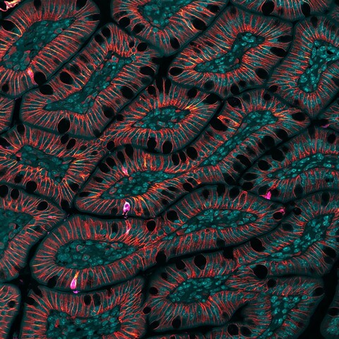
127 followers · 18 posts · Server mstdn.scienceImpaired growth factor signaling alters the cellular differentiation pattern of the small intestine #immunofluorescence #microscopy
#Immunofluorescence #microscopy
Mike Schumacher PhD · @schumacher
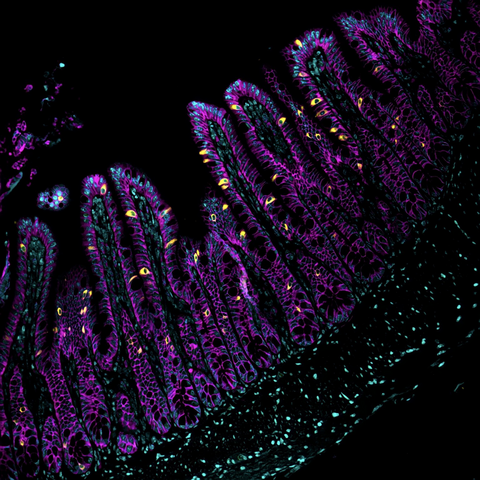
127 followers · 18 posts · Server mstdn.scienceTuft cells speckling the small intestine
#sciart #Immunofluorescence #microscopy
Mike Schumacher PhD · @schumacher
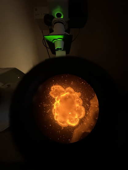
127 followers · 18 posts · Server mstdn.scienceWhat’s better than looking down the #microscope at a new fiercely bright antibody that worked?
#organoids #sciart #immunofluorescence #confocal #microscopy #gi
#microscope #organoids #sciart #Immunofluorescence #confocal #microscopy #gi
Mike Schumacher PhD · @schumacher
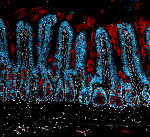
127 followers · 18 posts · Server mstdn.scienceIntestinal crypts filled with goblet cells—I love how in these #immunofluorescence sections you can see the mucus secreted into the crypt and intestinal lumen #sciart #microscopy #histology
#Immunofluorescence #sciart #microscopy #histology
Mike Schumacher PhD · @schumacher
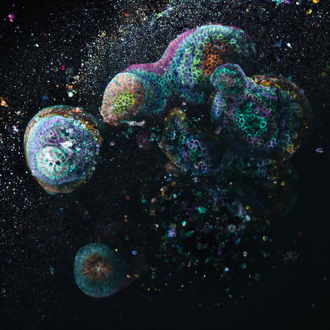
127 followers · 18 posts · Server mstdn.scienceA mini-gut intestinal #organoid imaged by #immunofluorescence confocal #microscopy ✨
#organoid #Immunofluorescence #microscopy #sciart #Science
Les Sutton · @LeslieSutton
57 followers · 79 posts · Server fediscience.orgToday for my #projects entry, I did some #microscopy work of some slides I've got! Things I learned:
- Mitoview green doesn't stick to mitochondria like antibodies do, the green WILL bleed into the cytoplasm after a few days. Image ASAP #immunocytochemistry
- been a minute since I did #confocalmicroscopy and forgot how much I loved it!
- maybe what I was thinking from my last experiment isn't true? Need more testing, more #Immunofluorescence and more confocal time!
#cancer #histology #ihc #Immunofluorescence #confocalmicroscopy #immunocytochemistry #microscopy #projects
Les Sutton · @LeslieSutton
57 followers · 79 posts · Server fediscience.orgSpending my morning doing some #confocal #microscopy, my lab usually uses our Echo Revolve for #immunofluorescence work and I've missed her so much 🥹
#Immunofluorescence #microscopy #confocal
Dr No & Mx Mo · @DrNoMxMo
84 followers · 38 posts · Server fediscience.orgDoes anyone know a good GPX4 #antibody for #WesternBlot and #Immunofluorescence reactive to humans and mice?
#Immunofluorescence #WesternBlot #antibody
