Katrina Velle · @KatrinaVelle
165 followers · 69 posts · Server mstdn.scienceBetter late than never for #MicroscopyMonday!
Here is a z-stack (every focal plane moving vertically through a sample) of actin polymer-stained Naegleria amoebae, followed by a maximum intensity projection (the brightest xy point of all the z positions). The darker colors here represent places where there is more actin polymer.
Katrina Velle · @KatrinaVelle
162 followers · 66 posts · Server mstdn.scienceHappy #MicroscopyMonday!
Here's a movie from the "Titus Takeover" week, where Meg Titus and several million Dictyostelium cells visited the Fritz-Laylin Lab!
Here are 2 movies (1st at 4x, 2nd at 10x) of Dicty cells streaming to form mounds during overnight starvation.
Mary Fesenko · @MAFesenko
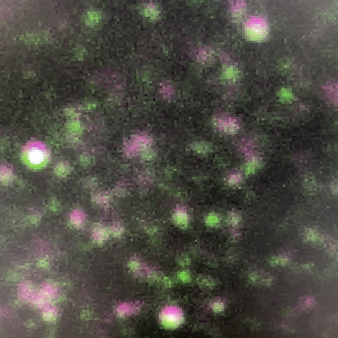
30 followers · 25 posts · Server mstdn.scienceSpent the afternoon in the #microscopy room, seeking meaning in patterns of multi-coloured dots again…sadly, falling prey to some wishful thinking along the way 🙃
Are you curious about what these pink and green dots are? Come to chat to @clathrin and I at the upcoming @J_Cell_Sci ‘Imaging Cell Dynamics’ meeting in Lisbon, where I will be presenting a poster!
#microscopy #MicroscopyMonday #cellbiology #celldyn2023
Katrina Velle · @KatrinaVelle
157 followers · 65 posts · Server mstdn.scienceHappy #MicroscopyMonday!
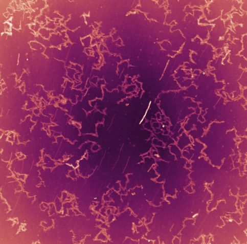
Here is a projection of 10 minutes of amoebae crawling on glass, so you can see their tracks. Not bad for a 4x objective and transmitted light!
LUT: CMOcean, curl
Katrina Velle · @KatrinaVelle
157 followers · 64 posts · Server mstdn.scienceHappy #MicroscopyMonday!
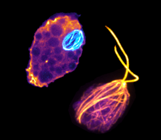
Here's Naegleria gruberi as a dividing amoeba and as a flagellate
Katrina Velle · @KatrinaVelle
156 followers · 63 posts · Server mstdn.scienceHappy #MicroscopyMonday!
Here is a ~very~ blebby amoeba making its way through a narrow channel.
Katrina Velle · @KatrinaVelle
156 followers · 63 posts · Server mstdn.scienceHappy #MicroscopyMonday!
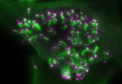
Here's one from my PhD work:
This cell is infected with Enteropathogenic E. coli (EPEC). EPEC translocates its own receptor (Tir, labeled pink) which triggers actin assembly (green) intro protrusive pedestals.
Katrina Velle · @KatrinaVelle
156 followers · 63 posts · Server mstdn.scienceHappy #microscopymonday!
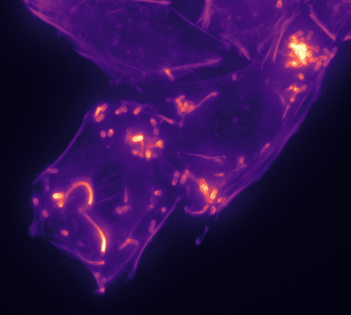
Here are some mammalian cells infected with Listeria, stained for actin polymer. ☄️
Katrina Velle · @KatrinaVelle
156 followers · 63 posts · Server mstdn.scienceHappy #MicroscopyMonday!
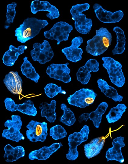
Here’s a collage I made a while back highlighting Naegleria’s actin (cyan) and microtubule (orange) cytoskeletons. Naegleria only use microtubules to build a mitotic spindle, and when they transiently transform into a flagellated cell type.
Katrina Velle · @KatrinaVelle
156 followers · 63 posts · Server mstdn.scienceHappy #MicroscopyMonday!
Here is an amoeba crawling through a microchannel. Blue areas show where the cell is in close contact with the coverslip (imaged by IRM), and DIC is shown in grayscale.
Elisabeth Kugler · @KuglerElisabeth
182 followers · 102 posts · Server mstdn.scienceBlood vessels of a developing #zebrafish.
Happy #MicroscopyMonday!
The vessels are about 10 micrometer in diameter. That's just a little bit bigger than blood cells ..
#zebrafish #MicroscopyMonday #Science #microscopy #vasculature
Katrina Velle · @KatrinaVelle
156 followers · 63 posts · Server mstdn.scienceHappy #MicroscopyMonday!
Here's a video of amoebae crawling around before and after the addition of a fluorescent membrane dye
Katrina Velle · @KatrinaVelle
156 followers · 63 posts · Server mstdn.scienceA little late for #MicroscopyMonday, but I still wanted to share this movie of Naegleria's contractile vacuoles that I just finished color coding!
Stacey Ogden 🦔 · @lab_ogden
245 followers · 93 posts · Server mstdn.scienceHere's a cute submission for #MicroscopyMonday ❤️ Shown is a primary cilium from an IMCD3 cell labeled with acetylated alpha-tubulin in red. Smoothened, the GPCR that is activated downstream of Sonic Hedgehog signals, is shown in green. To make the heart, the cilium image was duplicated and flipped using Photoshop. Have a great week!
Katrina Velle · @KatrinaVelle
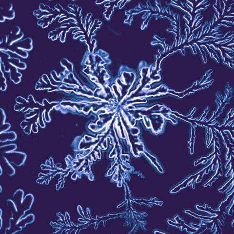
156 followers · 63 posts · Server mstdn.scienceHappy #MicroscopyMonday! Here is a crystal that formed in dried stabilized bleach.
Katrina Velle · @KatrinaVelle
156 followers · 63 posts · Server mstdn.scienceHappy #MicroscopyMonday!
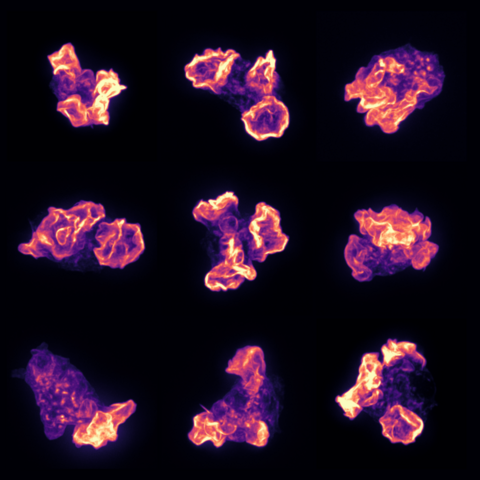
Here are some very ruffled amoebae, stained for actin polymer
LUT: mpl-magma
KaylinQ · @KaylinQ
23 followers · 80 posts · Server mstdn.science"It's #MicroscopyMonday, so let's take a dive under the  and check out muscle circulation of the adult amphipod (Parhyale hawaiensis), sometimes called the "sand flea." Credit: Longhua Guo. Taken during an MBL Embryology course, imaged on
@zeiss_micro
LSM 780."
https://twitter.com/MBLScience/status/1597296540179128322?s=20&t=uk8i9cEU32eaiG7voAgq8g
AIneurolab · @AIneurolab
16 followers · 6 posts · Server mstdn.scienceFor #MicroscopyMonday a Wnt1cre; tdtomato fetal mouse identifying dorsal root ganglia & craniofacial nerves and cartilages #neuralcrest #DevBio . Taken with a Zeiss Acmxio Zoom stereomicroscope.
#MicroscopyMonday #neuralcrest #DevBio
Spirochrome · @Spirochrome
92 followers · 6 posts · Server mstdn.scienceHoang Anh Le (aka Anh), PhD · @AnhHLe2702
156 followers · 48 posts · Server mstdn.scienceRT @AnhHLe2702
This cell has particularly huge mitochondria. You can see their cristae (you can see the zoom-in of the square area), which is the first time for me to ever see them so clearly! :O A belated #MicroscopyMonday #Fluorescence
#MicroscopyMonday #fluorescence
