CellBioNews · @cellbionews
90 followers · 1715 posts · Server scientificnetwork.deExploring potential of #periplasmic #biosynthesis for efficient solar-driven chemical production.
#bacteria #biomineralization #photosynthesis
https://phys.org/news/2023-07-exploring-potential-periplasmic-biosynthesis-efficient.html
#periplasmic #biosynthesis #bacteria #biomineralization #photosynthesis
Furqan Shah · @furqanshah
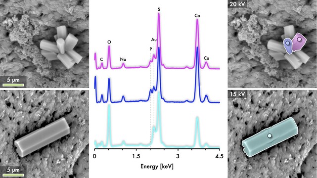
53 followers · 88 posts · Server mstdn.science🌸 Flower power – #calcium sulphate #crystals come in all shapes and forms! 💎🔬🔎🦴
#microscopy #electronmicroscopy #imaging #photography #biomineralization #biomaterials #sulphur #sulfur #crystallization #nature
#calcium #Crystals #microscopy #ElectronMicroscopy #imaging #photography #biomineralization #biomaterials #sulphur #sulfur #Crystallization #nature
Furqan Shah · @furqanshah
53 followers · 86 posts · Server mstdn.scienceMysterious #crystals on #bone exposed to tryptic soy broth: The plot thickens! 💎🦴
Elemental composition analysis using energy dispersive X-ray spectroscopy (#EDX/#EDS) picks up #calcium, #sulphur (#sulfur), but #phosphorus only occasionally! 🧪🔍
The #mystery crystals turn out to be calcium sulphate, with uncanny resemblance to the crystal habit of α-calcium sulphate hemihydrate!!!!! 🔬
Read the backstory here:
https://mstdn.science/@furqanshah/110020946034851733
#Crystals #bone #edx #calcium #sulphur #sulfur #phosphorus #mystery #Science #microscopy #biomineralization #elementalanalysis
Furqan Shah · @furqanshah
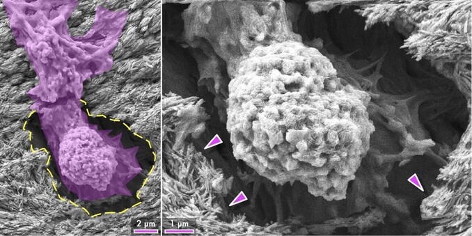
51 followers · 85 posts · Server mstdn.scienceAlien invasion!!! 👾 👽 Under in vitro culture, a #human #mesenchymal #stemcell crawls into an #osteocyte lacuna (http://bit.ly/Osteocyte) at the #bone surface, observed by scanning #electron #microscopy 🔬 🦴 Forget shape-memory #alloys! #Cells remember! (Arrowheads = bone #mineral)
#biology #electronmicroscopy #stemcells #imaging #bioimaging #biomimicry #biomineralization #invitro #apatite
#human #mesenchymal #stemcell #osteocyte #bone #electron #microscopy #alloys #cells #mineral #biology #ElectronMicroscopy #stemcells #imaging #bioimaging #biomimicry #biomineralization #invitro #apatite
Furqan Shah · @furqanshah
51 followers · 78 posts · Server mstdn.science#Magnesium #whitlockite formation in the alveolar #bone, with #bisphosphonate exposure and osteolytic #skeletal #metastasis. Scanning electron microscopy, Micro-Raman spectroscopy, and energy dispersive X-ray spectroscopy reveal Mg-rich, rhomboidal nodules (200 nm to 2.4 µm) within the lacuno-canalicular space. Mg-whitlockite formation in #osteocyte lacunae is multifactorial in #nature and suggests altered bone #biomineralization.
#magnesium #whitlockite #bone #bisphosphonate #skeletal #metastasis #osteocyte #nature #biomineralization #biology #pathology #Science
Furqan Shah · @furqanshah
45 followers · 73 posts · Server mstdn.scienceThrough the eyes of an #osteocyte. In #bone, this is what the environment of partially embedded osteocytes (osteoblastic–osteocytes) is like!
Scanning electron microscopy has been used to observe the orientation of mineral platelets in relation to osteoblastic–osteocyte lacunae on the surface of deproteinized trabecular bone in adult sheep.
Read more:
https://doi.org/10.1007/s00223-015-0072-8
@zeiss_micro #microscopy #biology #imaging #biomineralization #extracellularmatrix #friday
#osteocyte #bone #microscopy #biology #imaging #biomineralization #extracellularmatrix #friday
Furqan Shah · @furqanshah
45 followers · 73 posts · Server mstdn.science#Magnesium #whitlockite formation in the alveolar #bone, with #bisphosphonate exposure and #osteolytic #skeletal #metastasis. Scanning electron microscopy, Micro-Raman spectroscopy, and energy dispersive X-ray spectroscopy revealed Mg-rich, rhomboidal nodules (~200 nm to ~2.4 µm) within the lacuno-canalicular space. Mg-whitlockite formation within #osteocyte lacunae is multifactorial in #nature and suggests altered bone #biomineralization.
#magnesium #whitlockite #bone #bisphosphonate #osteolytic #skeletal #metastasis #osteocyte #nature #biomineralization #biology #pathology


