
preLights · @preLights
581 followers · 20 posts · Server biologists.social
A novel form of neuronal plasticity - when a neuron is injured, their healthy neighbour takes up the slack. 🗑️
First preLight from Matthew Davies (@sebastianrumpf) covering work from the #CarrilloLab
#preLight 👉 https://prelights.biologists.com/highlights/glial-draper-signaling-triggers-cross-neuron-plasticity-in-bystander-neurons-after-neuronal-cell-death/
#carrillolab #prelight #neuroscience #plasticity #celldeath #neurodegeneration

PLOS Biology · @PLOSBiology
4966 followers · 1603 posts · Server fediscience.org
Bak is a critical Bcl-2 family executor of #apoptosis. Structural, biochemical & cellular analyses reveal how Pxt1, a male #GermCell-specific protein, recognizes Bak to activate its pro-apoptotic activity and induce #CellDeath #PLOSBiology https://plos.io/43HVjQb
#plosbiology #celldeath #germcell #apoptosis

Phys.org · @physorg_bot
486 followers · 13947 posts · Server social.platypush.techReferenced link: https://phys.org/news/2023-06-protein-cell-membrane-rupture-death.html
Discuss on https://discu.eu/q/https://phys.org/news/2023-06-protein-cell-membrane-rupture-death.html
Originally posted by Phys.org / @physorg_com: http://nitter.platypush.tech/physorg_com/status/1665712297027747841#m
Protein serves as a breaking point for #cellmembrane rupture during #celldeath @unibasel @nature https://www.nature.com/articles/s41586-023-05991-z https://phys.org/news/2023-06-protein-cell-membrane-rupture-death.html

Claire McCarthy · @clairem402
154 followers · 283 posts · Server mstdn.social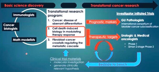
#ICYMI the recording of the #CancerMoonshot Seminar with Dr. Keith Syson Chan sharing how #celldeath induces non-classical mechanisms of #therapyresistance can be viewed at https://youtu.be/Nim2JWD9YkQ.
#cmsschan #ThrowBackThursday #therapyresistance #celldeath #cancermoonshot #icymi
PLOS Biology · @PLOSBiology
4896 followers · 1531 posts · Server fediscience.org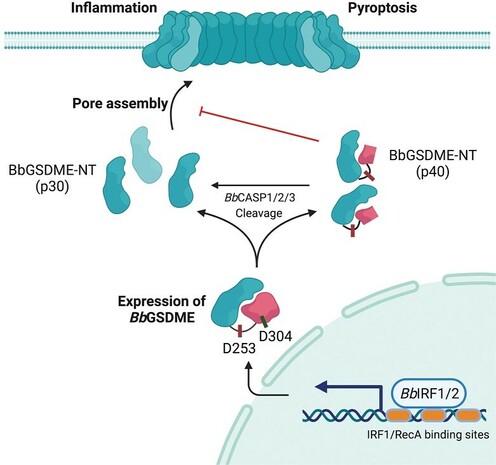
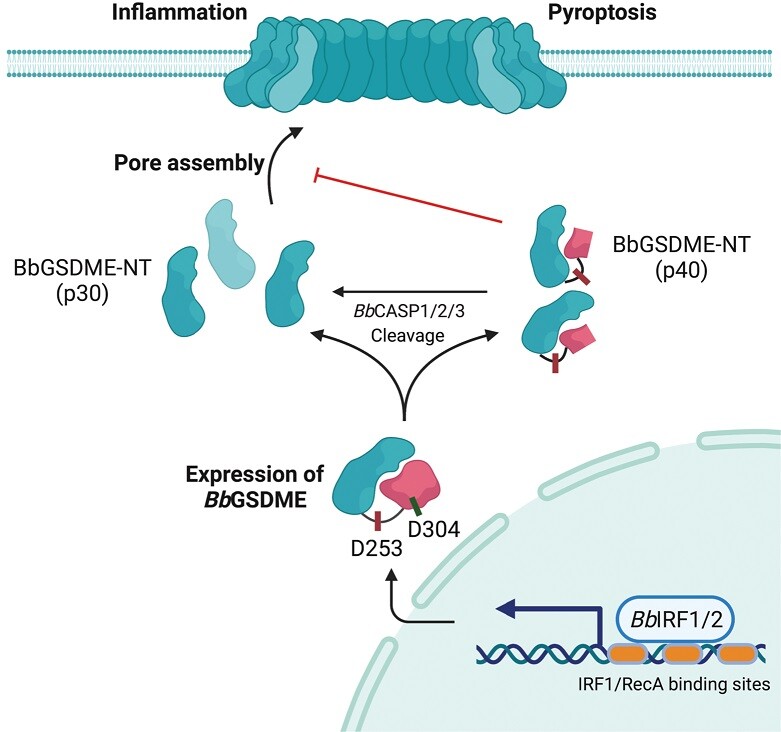
#Gasdermins (GSDMs) are pore-forming proteins involved in #CellDeath & #inflammation. @jianbin_ruan explores a #PLOSBiology study of the evolution of GSDMs in metazoa, highlighting the role of GSDME in #pyroptosis. Primer: https://plos.io/3nyd4By Paper: https://plos.io/3p6klcm
#pyroptosis #plosbiology #inflammation #celldeath #gasdermins
PLOS Biology · @PLOSBiology
4897 followers · 1529 posts · Server fediscience.org
#Gasdermins (GSDMs) are pore-forming proteins involved in #CellDeath & #inflammation. @jianbin_ruan explores a #PLOSBiology study of the evolution of GSDMs in metazoa, highlighting the role of GSDME in #pyroptosis. Primer: https://plos.io/3nyd4By Paper: https://plos.io/3p6klcm
#pyroptosis #plosbiology #inflammation #celldeath #gasdermins
PLOS Biology · @PLOSBiology
4897 followers · 1524 posts · Server fediscience.org
#Gasdermins (GSDMs) are pore-forming proteins involved in #CellDeath & #inflammation. @jianbin_ruan explores a #PLOSBiology study of the evolution of GSDMs in metazoa, highlighting the role of GSDME in #pyroptosis. Primer: https://plos.io/3nyd4By Paper: https://plos.io/3p6klcm
#pyroptosis #plosbiology #inflammation #celldeath #gasdermins

Thiago Carvalho · @cyrilpedia
1640 followers · 6928 posts · Server qoto.orgA long & winding road.
"However, our efforts to identify a chemical inhibitor of apoptosis floundered. Whatever we tested either did not work or had only a marginal effect. We wondered whether components of the pathway might have built-in redundancies. If so, the project was dead."
#Apoptosis #CellDeath
https://www.science.org/doi/10.1126/sciadv.adi2011#.ZEmbzTtPNQI.twitter
Phys.org · @physorg_bot
438 followers · 11142 posts · Server social.platypush.techReferenced link: https://phys.org/news/2023-04-scientists-reveal-ferroptosis-cell-death.html
Discuss on https://discu.eu/q/https://phys.org/news/2023-04-scientists-reveal-ferroptosis-cell-death.html
Originally posted by Phys.org / @physorg_com: http://nitter.platypush.tech/physorg_com/status/1643661973798649870#m
Scientists reveal more about the process behind ferroptosis, a recently discovered type of #celldeath @tohoku_univ @currentbiology https://linkinghub.elsevier.com/retrieve/pii/S0960982223002002 https://phys.org/news/2023-04-scientists-reveal-ferroptosis-cell-death.html

stjuderesearch · @stjuderesearch
19 followers · 47 posts · Server ohai.social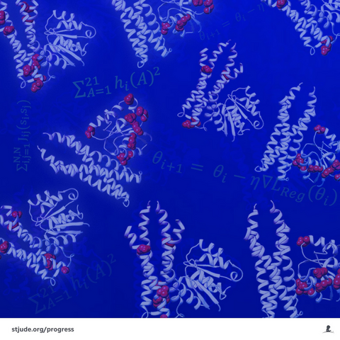
Researchers at #StJude have developed an #algorithm inspired by #evolutionary principles to #design new #protein pairs that interact exclusively. With this approach, it might be possible, for example, to design proteins or short #peptides that can inhibit or activate #celldeath proteins, which would be useful to help treat a variety of #metabolicdiseases. Learn more. https://bit.ly/3ZBy9sr
#StJudeResearch #DataScience
#stjude #algorithm #evolutionary #design #protein #peptides #celldeath #metabolicdiseases #stjuderesearch #datascience
PLOS Biology · @PLOSBiology
4726 followers · 868 posts · Server fediscience.org
The past 20 years have revealed several new #InnateImmune sensing & #CellDeath pathways with disease relevance. @RebeccaTweedell @KannegantiLab look back at the impact of these discoveries & forwards toward their future therapeutic potential #PLOSBiology https://plos.io/40O9xxQ
#plosbiology #celldeath #innateimmune
PLOS Biology · @PLOSBiology
4727 followers · 865 posts · Server fediscience.org
The past 20 years have revealed several new #InnateImmune sensing & #CellDeath pathways with disease relevance. @RebeccaTweedell @KannegantiLab look back at the impact of these discoveries & forwards toward their future therapeutic potential #PLOSBiology https://plos.io/40O9xxQ
#plosbiology #celldeath #innateimmune
PLOS Biology · @PLOSBiology
4724 followers · 860 posts · Server fediscience.org
The past 20 years have revealed several new #InnateImmune sensing & #CellDeath pathways with disease relevance. @RebeccaTweedell @KannegantiLab look back at the impact of these discoveries & forwards toward their future therapeutic potential #PLOSBiology https://plos.io/40O9xxQ
#plosbiology #celldeath #innateimmune
stjuderesearch · @stjuderesearch
17 followers · 26 posts · Server ohai.social
Thirumala-Devi Kanneganti, PhD, has been named a Fellow of the American Association for the Advancement of Science for achievements in innate immunity, developing new concepts in inflammasome biology and identifying an inflammatory cell-death pathway called PANoptosis. https://bit.ly/Kanneganti-AAAS
#StJude #StJudeResearch #Immunology #WomenInSTEM #CellDeath
#stjude #stjuderesearch #immunology #womeninstem #celldeath
Thiago Carvalho · @cyrilpedia
1494 followers · 4248 posts · Server qoto.org
"Our findings underscore the importance of cell death in defense against intracellular bacterial pathogens and provide an example of how layered and hierarchical immune pathways can provide robust defense against pathogens that have evolved a broad arsenal of virulence factors."
#Shigella #HostPathogen #CellDeath #Apoptosis #InnateImmunity #Pathogen #Microbiology #Immunology
#apoptosis #pathogen #shigella #hostpathogen #celldeath #microbiology #innateimmunity #immunology

The EMBO Journal · @embojournal
387 followers · 74 posts · Server sciencemastodon.com
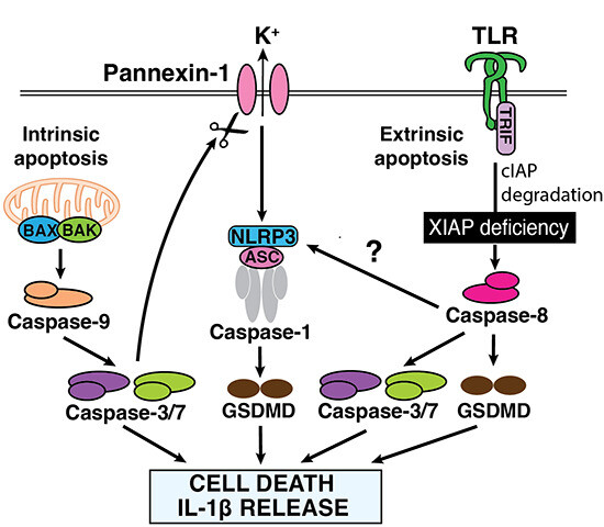
#Caspase8-driven #apoptotic and #pyroptotic crosstalk causes #celldeath and #interleukin IL-1β release in X-linked inhibitor of apoptosis (#XIAP) deficiency
#caspase8 #apoptotic #pyroptotic #celldeath #interleukin #xiap

Joseph P. · @tonic
137 followers · 344 posts · Server qoto.orgHow Mitochondrial Dynamism Orchestrates Mitophagy
Authors Orian S. Shirihai, Moshi Song, Gerald W. Dorn II
Understanding the Significance of Mitochondrial Fission and Fusion
Mitochondrial dynamics refers to the movement of #mitochondria within a cell. This includes #fission, which is when mitochondria divide into two parts, #fusion, which is when two mitochondria join together, and #translocation, which is when mitochondria move from one part of the #cell to another. This movement is important for maintaining the stability of the mitochondrial #DNA, which is the genetic material found in mitochondria, and for controlling the cell's #respiration. It can also be involved in programmed #CellDeath. In the #heart, mitochondrial dynamics #protein s, such as #mitofusin s, optic #atrophy, and dynamin-related protein, are highly expressed and play an important role in maintaining the quality of the #mitochondria. Other roles for mitochondrial dynamics proteins in the #heart include helping to move #calcium into the mitochondria and regulating the structure of the mitochondria.
#Mitochondria are organelles in cells that are responsible for producing #energy. They can change their structure by breaking apart (#fission) and reforming (#fusion). This process is complicated and energy intensive, so it is important to understand why it is necessary. One reason may be that when cells divide, the mitochondria need to be divided equally between the two daughter cells. This requires the #mitochondria to be broken apart and then reformed in each daughter #cell. This process of breaking apart and reforming is more efficient than growing and budding the mitochondria. To help explain this process, the authors use the analogy of an army. Each soldier in the army is like a protein in the mitochondria, and the different units of the army are like the different parts of the #mitochondria. To increase the size of the army, units are added, rather than individual soldiers. This is similar to how mitochondria are modified, by adding or subtracting intact functional units, rather than individual #protein s.
Mitochondria are #organelle s in cells that can change their physical structure by undergoing fission. Fission can be symmetrical, which is when mitochondria replicate and expand the number of #mitochondria in the #cell, or asymmetrical, which is when damaged components of the mitochondria are removed. The major #protein that helps with mitochondrial fission is called Drp1. It is mostly found in the #cytosol, but it needs to be recruited to the outer mitochondrial #membrane to help with fission. Different factors can cause Drp1 to be recruited, such as phosphorylation by mitotic kinase cyclin B–cyclin-dependent-kinase (cdk) 1 complex during #cell division, or interacting with Bcl-2–associated protein x during #apoptosis. Inhibiting Drp1 can protect cells from some, but not all, forms of programmed cell death.
Mitochondria, which are organelles in cells, can be partitioned in #mitosis. The most efficient way to do this is by dismantling and then reconstituting the cellular #mitochondria network through sequential #organelle fission, distribution, and refusion. To explain this concept, the text uses an analogy of how military units are constituted and managed within an army's hierarchical organization structure. In this analogy, each soldier represents an individual respiratory complex #protein, which are grouped together to form a squad (analogous to a respiratory complex). Squads are arranged into platoons, and approximately 6 platoons comprise a functional unit, the company (like 1 complete respiratory chain). The text suggests that it would be easier to add prefabricated supercomplexes to preexisting ones, as by fusing mitochondrial cristae, rather than trying to make a larger or different shaped mitochondrion through the wholesale incorporation of individual proteins. This is because making major structural modifications of respiratory supercomplexes on paracrystalline cristal membranes would first require destabilizing the #membrane, then incorporating additional individual #protein components, and finally reconstructing the original highly organized structure, which is complicated and potentially disruptive.
#Mitochondria are small organelles in cells that can change their physical structure by undergoing fission. Fission can be symmetrical, which means the #mitochondria are split into two equal parts, or asymmetrical, which means the mitochondria are split into two unequal parts. Symmetrical fission is used to replicate and expand the number of mitochondria in the #cell, while asymmetrical fission is used to remove damaged mitochondria from the cell. The major #protein responsible for mitochondrial fission is called Drp1. Drp1 is mostly found in the cytosol, but it needs to be recruited to the outer mitochondrial #membrane to promote fission. Different factors can stimulate Drp1 to move to the outer mitochondrial #membrane, such as phosphorylation by mitotic kinase cyclin B–cyclin-dependent-kinase (cdk) 1 complex. In addition, the endoplasmic reticulum (ER) is often found at the sites of mitochondrial fission. If Drp1 is not present, the mitochondria can still fragment during #mitosis, suggesting that there are other mechanisms that can promote mitochondrial fission.
The text is talking about the process of mitochondrial fission, which is a process that involves connecting and separating parts of a #mitochondria. The author uses the metaphor of making sausage links to explain the process, but then goes on to explain that mitochondria are actually more like a turducken, which is a dish made of a chicken stuffed inside a duck stuffed inside a turkey. This creates layers of poultry, which is similar to the double #membrane /double space structure of #mitochondria. The author then explains that the process of mitochondrial fusion involves connecting the two mitochondria layer by layer, using proteins called mitofusins. Mitofusins have a #GTPase domain, two hydrophobic heptad repeat coiled-coil domains, and a small hydrophobic transmembrane domain. These proteins insert into the outer #membrane of the #mitochondria, and can interact with other proteins in the cytosol. The process of mitochondrial fusion is GTP-independent and reversible, but #GTP #hydrolysis is essential for irreversible outer membrane fusion.
#Mitofusins are proteins that are essential for the first two stages of mitochondrial fusion, which is the process of two mitochondria joining together. This process is important for the exchange of information between the #mitochondria and the #cell. If the mitofusins are deleted or suppressed, the mitochondria become abnormally small and are unable to undergo normal fusion. This can have serious implications for the health of the #cell.
Membrane-by-membrane mitochondrial fusion is a process that helps to keep the structure of the inner and outer membranes of #mitochondria intact. This helps to preserve the process of oxidative phosphorylation, which is important for providing energy to cells. Without this process, molecules that can be toxic to cells can form and interrupt the electron transport chain. This process is also important for maintaining the normal shape of the crista, which is necessary for the proper assembly and functioning of electron transport chain supercomplexes. In addition, it has been shown that interrupting Mfn-mediated OMM fusion can cause a #cardiomyocyte ER stress response, while interrupting Opa1-mediated IMM fusion can compromise mitochondrial function.
Mitochondrial fission and fusion are important processes in #biology, as evidenced by the fact that mutations in genes related to these processes can cause serious diseases in humans. Altering the balance between fission and fusion can have an effect on the shape of #mitochondria, with more fusion leading to longer, more interconnected mitochondria, and more fission leading to shorter, less interconnected mitochondria. It is generally thought that more interconnected #mitochondria are healthier, but this is not always the case. In some cases, mitochondrial #fragmentation can be beneficial, and it is important to understand the interplay between mitochondrial fragmentation and other processes, such as #mitophagy, in order to understand the effects of mitochondrial fission and fusion.
Mitophagy is a process by which cells eat their own #mitochondria. Mitochondria are organelles that produce energy in the form of #ATP, which is used to power most biological processes. Over time, mitochondria can become damaged and produce toxic levels of reactive oxygen species ( #ROS ). To protect the #cell from this damage, it has developed a sophisticated system to identify and remove these dysfunctional #mitochondria. This process is called mitophagy. #Mitophagy is a combination of the words mitochondria and #autophagy, which means "self-eating". It is a way for cells to selectively target and remove damaged mitochondria, while still keeping healthy ones. This helps to maintain the balance between having enough energy-producing #mitochondria and getting rid of the ones that are no longer functioning properly.
Pulse chase experiments are a type of scientific experiment used to study the behavior of molecules over time. In this particular experiment, researchers found that when #mitochondria (the energy-producing organelles in cells) are targeted for #mitophagy (a process of removing damaged mitochondria from the cell), they have a relatively depolarized #membrane potential before being removed. This means that the #mitochondria have a lower electrical charge than normal, and they are less likely to be involved in #fusion events (when two mitochondria join together). The time between the mitochondria becoming depolarized and being removed from the cell can range from less than an hour to about three hours, suggesting that there is a population of preautophagic #mitochondria (mitochondria that are about to be removed). This #preautophagic pool helps to explain the variation in mitochondrial #membrane potential in different cell types. The process that feeds mitochondria into the preautophagic pool is important for determining how quickly #mitochondria are removed from the #cell. Scientists have developed a technology to label individual mitochondria and track their #membrane potential, which has allowed them to identify the event at which depolarized #mitochondria are produced. This event is called asymmetrical fission, and it occurs when the daughter mitochondria produced by the fission event have different #membrane potentials - one daughter has a higher membrane potential than the mother mitochondrion, while the other daughter has a lower membrane potential. This process of asymmetrical fission helps to separate damaged components from healthy components before they are removed from the #cell.
The concept of mitochondrial fission and fusion and how it affects mitochondrial quality. It suggests that when the fusion factors Mfn1 and Mfn2 are both absent, unusually small and degenerated #mitochondria accumulate in adult mouse hearts. This was associated with impaired #cardiomyocyte respiration, but not with measurable alterations in #oxygen consumption. It was later discovered that the isolation procedure used was not capturing the fragmented #mitochondria produced by interrupting mitochondrial fusion. This led to the discovery that Mfn2 is essential to #Parkin-mediated #mitophagy, which is a process that helps to maintain mitochondrial quality. Three recent papers have also implicated the mitochondrial fission protein Drp1 in cardiac #mitophagy, and it is suggested that if asymmetrical mitochondrial fission normally precedes mitophagy, then chronic suppression of fission by ablating Drp1 would have different consequences on #mitophagy depending on when it is assayed.
Mfn2 and PINK1–Parkin Mitophagy Signaling is a mechanism for controlling the quality of #mitochondria in the body. #PINK1 and #Parkin are proteins that are linked to #Parkinson's disease, and mutations in their genes were the first to be identified as causing the disease. Scientists have studied how PINK1 interacts with Parkin, and how this interaction can lead to the destruction of damaged #mitochondria, which is called #mitophagy. #PINK1 is like an ignition switch that senses when mitochondrial damage has occurred, and then activates Parkin-mediated mitophagy. PINK1 is normally not present in healthy #mitochondria, but when mitochondrial damage occurs, PINK1 accumulates and triggers the destruction of the damaged #mitochondria.
PINK1 is a protein that accumulates on damaged mitochondria and helps to promote mitophagy, which is the process of getting rid of damaged mitochondria. PINK1 does this by inducing the cytosolic protein Parkin to move to the mitochondria and ubiquitinate proteins on the outer membrane of the mitochondria. This helps to prevent the spread of damage from the damaged mitochondria to the healthy ones. PINK1 also inhibits the fusion of the damaged mitochondria. There are different theories about the biochemical events that cause Parkin to move to the mitochondria and stop the fusion. It is thought that PINK1 phosphorylates Parkin on certain sites, which helps Parkin bind to the mitochondria. It is also thought that PINK1 phosphorylates ubiquitin, which helps Parkin bind to the mitochondria and ubiquitinate proteins on the outer membrane. Finally, it is thought that PINK1 phosphorylates Mfn2, which helps Parkin bind to the mitochondria and ubiquitinate proteins on the outer membrane. All of these processes help to promote mitophagy and prevent the spread of damage from the damaged mitochondria to the healthy ones.
#PINK1 is a protein that plays an important role in a process called #mitophagy, which is a form of quality control for mitochondria. Mutations in the #PINK1 #gene have been linked to hereditary #Parkinson's disease in humans, but when the PINK1 gene is deleted in mice, it does not cause the same #neurodegenerative pattern seen in humans. Even when the genes for PINK1, Parkin, and DJ-1 are all deleted in mice, it still does not cause the same loss of dopaminergic #neuron s seen in #Parkinson's disease patients. This suggests that there may be other pathways that can compensate for the loss of #PINK1 and #Parkin, such as increased transcription of other E3 #ubiquitin ligases in the hearts of Parkin-knockout mice.
The text is discussing the idea of mitochondrial quality control pathways, which are processes that help keep mitochondria healthy. The text is suggesting that there may be alternate pathways that can be used to maintain mitochondrial health, rather than waiting until the mitochondria are completely depolarized before triggering their removal. It is comparing this idea to the idea of maintaining a car, where it is better to perform regular maintenance and repairs rather than waiting until the car is completely broken down before replacing it.
Like a car, mitochondria can be maintained through preventative maintenance, such as replacing worn parts, and that more serious damage can be repaired by removing and replacing individual components. It also suggests that, like a car, #mitochondria can be repaired by removing and replacing damaged parts, but on a smaller scale. The different types of maintenance and repair may be part of a continuum, rather than distinct categories.
#Mitophagy and mitochondrial dynamism are two processes that are closely connected. Mitophagy is the process of removing damaged #mitochondria from the #cell, while mitochondrial dynamism is the process of mitochondria fusing together and separating. The two processes work together to keep the cell healthy by eliminating damaged mitochondria and preventing healthy mitochondria from being contaminated by the damaged ones. The protein #Mfn2 plays a role in both processes, acting as a factor for mitochondrial fusion when it is not acted on by #PINK1 and as a receptor for #Parkin when it is. This suggests that the two processes are mutually exclusive, meaning that they cannot happen at the same time. This helps to protect healthy #mitochondria from being contaminated by the damaged ones. Finally, the involvement of PINK1 and Parkin in multiple mitochondrial quality control mechanisms shows that there are multiple ways to keep the #mitochondria healthy, which is important for preventing chronic degenerative diseases and providing opportunities for #therapeutic intervention.
#dna #mitosis #cardiomyocyte #biology #autophagy #oxygen #parkin #celldeath #atrophy #calcium #cytosol #fusion #explainpaper #fission #translocation #protein #mitochondria #cell #respiration #heart #mitofusin #energy #organelle #gtpase #gtp #membrane #hydrolysis #Mitofusins #fragmentation #apoptosis #mitophagy #atp #ros #preautophagic #PINK1 #parkinson #gene #neurodegenerative #neuron #ubiquitin #Mfn2 #therapeutic

Kurianlab · @kurianlab
45 followers · 104 posts · Server sciencemastodon.comGives me a lot of pleasure to share this from my talented, clever collegue and neighbour @ale_annibaldi81. New insights into how c-flip cleavage acts as a decision point between cell death and regenerative response. Please RT
---
RT @ale_annibaldi81
Delighted to share the #AnnibaldiLab first Preprint. We found a previously unrecognized function for #cFlip cleavage in limiting #CellDeath during viral infection and injury, to promot…
https://twitter.com/ale_annibaldi81/status/1603767171007627264
#annibaldilab #cflip #celldeath
Joseph P. · @tonic
134 followers · 288 posts · Server qoto.org
Tumoral Immune Cell Exploitation in Colorectal Cancer Metastases Can Be Targeted Effectively by Anti-CCR5 Therapy in Cancer Patients
Niels Halama, Inka Zoernig, Anna Berthel, Christoph Kahlert, Fee Klupp, Meggy Suarez-Carmona,Thomas Suetterlin, Karsten Brand, Juergen Krauss, Felix Lasitschka, Tina Lerchl, Claudia Luckner-Minden, Alexis Ulrich, Moritz Koch, Juergen Weitz, Martin Schneider, Markus W. Buechler, Laurence Zitvogel,
Thomas Herrmann, Axel Benner, Christina Kunz, Stephan Luecke, Christoph Springfeld, Niels Grabe, Christine S. Falk, and Dirk Jaeger
Targeting Tumor-Promoting Microenvironment Through CCR5 Blockade in #Colorectal #Cancer #Liver Metastases
#Cancer progression is a process in which cancer cells and #immune cells interact with each other in a way that can lead to the growth and spread of cancer. In #colorectal cancer, when the cancer has spread to other parts of the body, it is called #metastasis and it is very difficult to treat. Treatments such as PD-1/PD-L1 blockade and #chemokine modulation have been successful in modifying the interactions between the immune system and cancer, leading to the rejection or suppression of progression. Cancer cells can also alter the immune microenvironment, leading to #immunosuppression and #immune evasion. In this research paper, the authors studied the microenvironment in #CRC #liver metastases and identified a network of #tumor cells and immune cells that exploit the CCL5-CCR5 axis. They then investigated and characterized the effects of blocking the CCL5-CCR5 axis.
the microenvironment of #liver metastases of #colorectal cancer (#CRC).
the environment induces migration of T lymphocytes, which produce a #cytokine called CCL5. This CCL5 then supports tumor growth and spread by influencing macrophages and #tumor cells. The environment is immunosuppressive and the tumor cells are exploiting the host's #immune cells to their advantage. In other words, the tumor cells are using the host's immune cells to help them grow and spread.
the effects of CCR5 blockade on the #tissue level.
Tumor #cell death and a specific pattern of #cytokine and #chemokine modulation are observed in the #ExplantModel and in #tumor biopsies from a #ClinicalTrial. Macrophages are the key for these anti-tumoral effects, as they produce IFNs and reactive oxygen species which cause tumor cell death. #CCR5 blockade induces a phenotypic shift in the macrophages, which is referred to as a switch from an M2 to an M1 phenotype. This repolarization also reduces levels of CD163+ cells, reshaping the #myeloid cell composition in the microenvironment. The influx of new effector cells due to CCR5 inhibition can shift the effects of CCL5 towards beneficial effects, such as reduction of #immunosuppression , #angiogenesis, and #chemotherapy resistance.
The microenvironment of the invasive margin of #liver metastases.
There was no relevant Th1, Th2, or Th17 #cytokine signature present in any of the samples. However, the authors did find that #chemokines and #macrophage-related cytokines were significantly increased at the invasive margin. Chemokines are molecules that help to attract #immune cells to the area, and macrophage-related cytokines are molecules that help to regulate the activity of #macrophages, which are a type of immune cell. 98% of the CD3+ #lymphocyte s in the resection specimens were positive for PD-1, which is a molecule that helps to regulate the activity of the immune system.
#CCL5 is a protein produced by T cells, which are a type of white blood cell. #CCR5 is a receptor found on metastatic tumor cells, which are cancer cells that have spread from the primary #tumor to other parts of the body. In this research paper, it was found that CCL5 has #pleiotropic tumor-promoting effects on #tumor cells and tumor-associated #macrophage s. This means that CCL5 has multiple effects on both the cancer cells and the macrophages, which are a type of white #blood #cell, that are associated with the #tumor. CCL5 was produced mainly by T cells located at the invasive margin and #peritumoral stroma of metastases, and that CCR5 was dominantly expressed by metastatic tumor cells. CCL5 also had effects on tumor #CellProliferation, invasive tumor #CellBehavior, and increased production of matrix #metalloproteinas es by tumor-associated macrophages. Finally, they found that CCR5 inhibition had an effect on key molecules of #epithelial to #mesenchymal transition ( #EMT ).
The researchers wanted to test the effects of #CCR5 blockade, which is a way of blocking the CCR5 receptor on cells, using a drug called maraviroc. They used human #tumor #explantmodel s, which are samples of #tissue from advanced #CRC patients with #liver metastases. Maraviroc led to morphologically overt tumor #CellDeath in the #explants, which means that the tumor cells died and changed in appearance. The researchers then tested the hypothesis that #macrophage s, (type of white blood cell), were required for the tumor cell death-inducing effects of CCR5 blockade. They used clodronate #liposome s to deplete CD163+ TAMs, ( #macrophage s associated with tumors) and found that combining clodronate with CCR5 inhibition abrogated the immediate tumor cell death-inducing effects of #CCR5 inhibition. This confirmed the role of macrophages in this process. IFN-g induced stromal CD163+ #macrophage #cell death and led to a reconfiguration of the #myeloid cell compartment. Inhibition of macrophage-derived reactive oxygen species could partially block the anti-tumoral effects of CCR5 inhibition. Finally, they tested the effects of CCL5/CCR5 inhibition and found that both a CCL5 neutralizing antibody and a CCR5 blocking #antibody had similar functional effects to maraviroc.
A #ClinicalTrial (MARACON) was conducted to test the effects of a drug called maraviroc on patients with advanced-stage #metastatic colorectal #cancer. The #trial involved taking biopsies of the patients before and after treatment with maraviroc, and the results showed that the drug had beneficial effects on the tumor-promoting #microenvironment and led to objective clinical responses. These responses included induction of central #TumorNecrosis, reduction of tumor cell death, and reduction of key #cytokine s and growth factors that promote tumor growth. The drug was also found to be very well tolerated, with mild elevation of #liver enzymes being the most common side effect. Finally, the trial showed that partial responses were achieved in patients with previously refractory disease.
CCR5 blockade, is a type of #therapy used to treat #cancer.
The MARACON clinical trial, showed that CCR5 blockade had a positive effect on the tumor microenvironment and led to a higher response rate in subsequent chemotherapies. The authors suggest that this effect is not limited to the #liver metastases, but is a systemic feature. They also suggest that the local presence of multiple layers of #immune subversion in cancers depends on the individual tissue, #treatment, tumor type, and the difference between primary #tumor and metastatic lesion. The authors also found that the results of the #ClinicalTrial were in line with the results of a fully human organotypic tumor #ExplantModel, which is a simple model with a straightforward approach. The authors also note that the survival data from the trial is not conclusive due to the limited number of patients, but that the objective treatment responses are very encouraging. They suggest that CCR5 blockade may be a promising approach and needs to be evaluated further scientifically and clinically.
#liver #immune #metastasis #chemokine #immunosuppression #crc #cytokine #tissue #cell #ExplantModel #clinicaltrial #ccr5 #myeloid #angiogenesis #chemotherapy #CellProliferation #CellBehavior #therapy #treatment #chemokines #macrophage #macrophages #lymphocyte #CCL5 #pleiotropic #blood #peritumoral #metalloproteinas #epithelial #mesenchymal #emt #celldeath #explants #liposome #antibody #metastatic #trial #microenvironment #TumorNecrosis #colorectal #cancer #tumor
Thiago Carvalho · @cyrilpedia
971 followers · 1762 posts · Server qoto.orgThirty years ago, in November 1992, Ishida, Honjo and their collaborators reported the discovery of the #PD1 gene in @embojournal
https://www.embopress.org/doi/abs/10.1002/j.1460-2075.1992.tb05481.x
I spoke with Tasuku Honjo, Pierre Golstein (whose team first cloned #CTLA4) & Facundo Batista about the discovery earlier this year.
The initial project had absolutely nothing to do with checkpoints or #immunotherapy
Listen to the full #EMBOPodcast episode at the link below or on pretty much any podcast app
#Immunology #HistoryScience #Cancer #CellDeath
https://www.embo.org/podcasts/from-cell-death-to-cancer-immunotherapy/
#pd1 #HistoryScience #cancer #celldeath #CTLA4 #immunotherapy #EMBOPodcast #immunology