
Christophe Leterrier 🔬 · @christlet
1309 followers · 346 posts · Server mas.to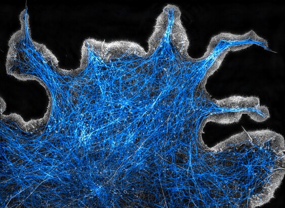
Spending time with a microscope, pushing it, calibrating it over and over, until it feels right #cellfie
Christophe Leterrier 🔬 · @christlet
1378 followers · 356 posts · Server mas.toSpending time with a microscope, pushing it, calibrating it over and over, until it feels right #cellfie
Christophe Leterrier 🔬 · @christlet
1305 followers · 343 posts · Server mas.to
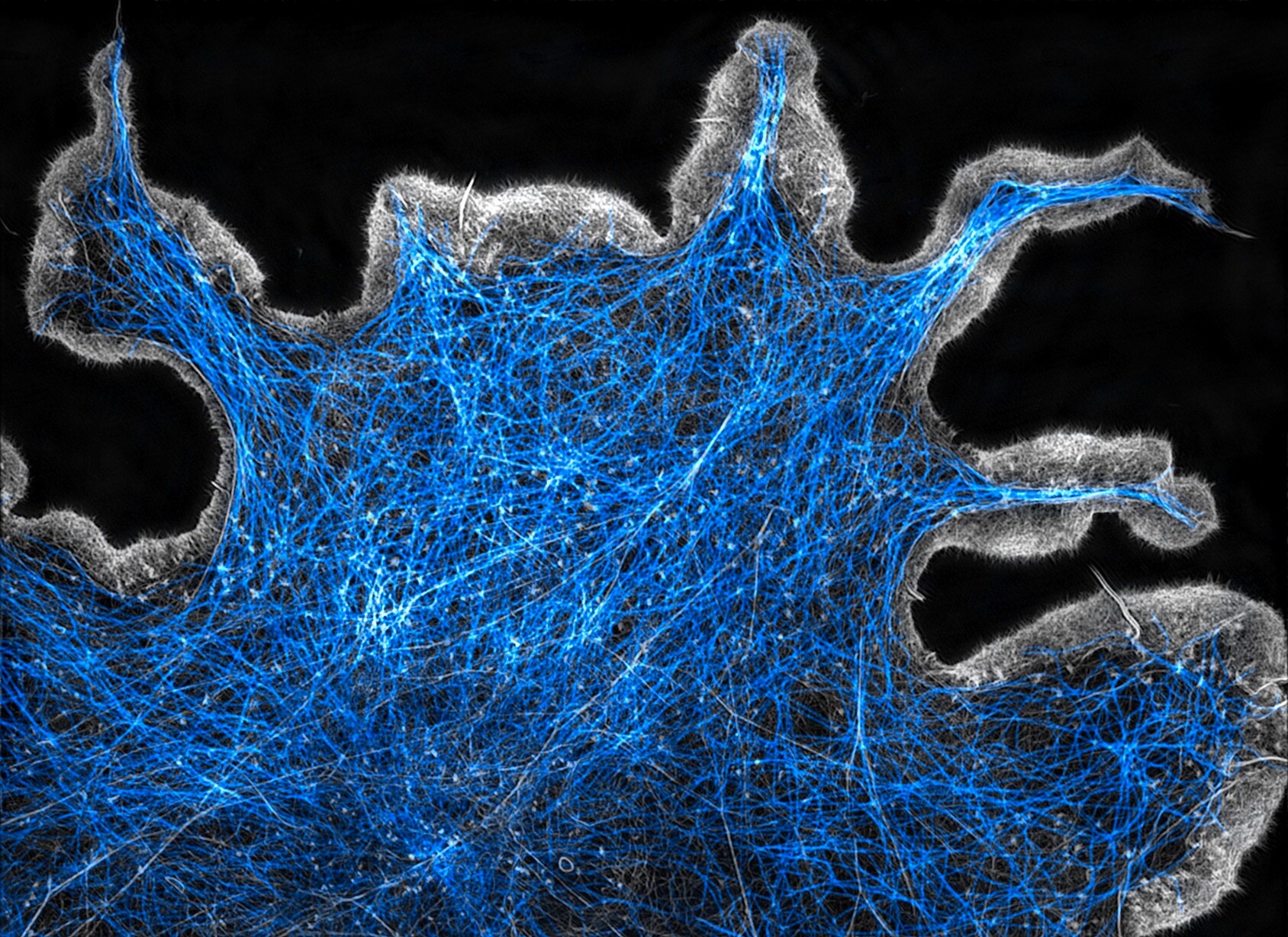
Struggled with SIM calibration in this freezing weather - the scope likes its toasty 23°C room temperature! But now it's OK #realtimemicroscopy #cellfie
Christophe Leterrier 🔬 · @christlet
1378 followers · 356 posts · Server mas.to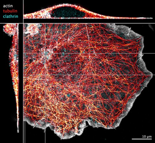
Struggled with SIM calibration in this freezing weather - the scope likes its toasty 23°C room temperature! But now it's OK #realtimemicroscopy #cellfie

Andrew Burgess · @AndrewBurgessPhD
205 followers · 154 posts · Server mastodon.social
Throwback Thursday. #microscope #cellfie A mouse embryo fibroblast #MEF stained for #microtubule (blue) and #DNA (green). Images taken on a #Leica #confocal #SP8
#microscope #cellfie #mef #microtubule #dna #leica #confocal #sp8

Leica Microsystems · @leicamicrosystems
50 followers · 10 posts · Server mastodon.social
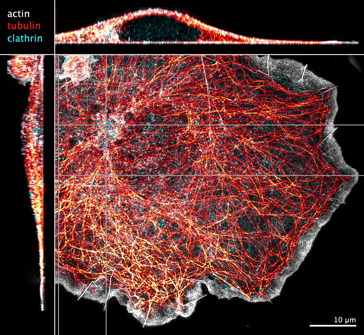
🔬 📸 Check out this image of cultured human retinal pericytes stained for vimentin (cyan), F-actin (magenta), and DAPI (blue). Giulia De Rossi uses in vitro culture systems to look at how LRG1 affects pericyte morphology and function. Imaged with a #STELLARIS 5 confocal microscope.
.
✅ More about STELLARIS:
https://fcld.ly/60v3ai9
#ScienceTwitter #ScienceMastodon #cellfie #cell #microscopy #science #research
#stellaris #sciencetwitter #sciencemastodon #cellfie #cell #microscopy #science #research
dgaboriau · @dgaboriau
105 followers · 11 posts · Server mas.to
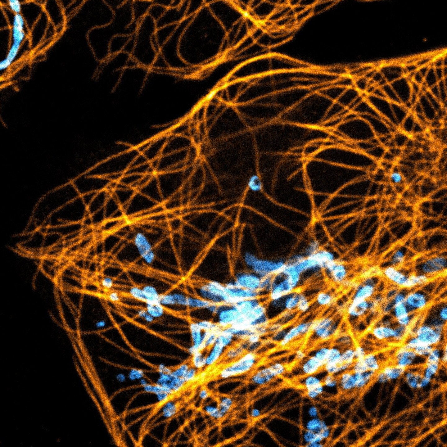
Another #cellfie, tubulin and mitochondria on @leicamicrosystems Stellaris 8 confocal

Christophe Leterrier · @christlet
439 followers · 42 posts · Server mas.to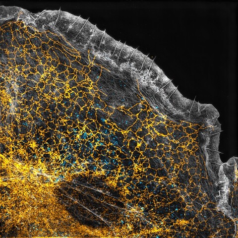
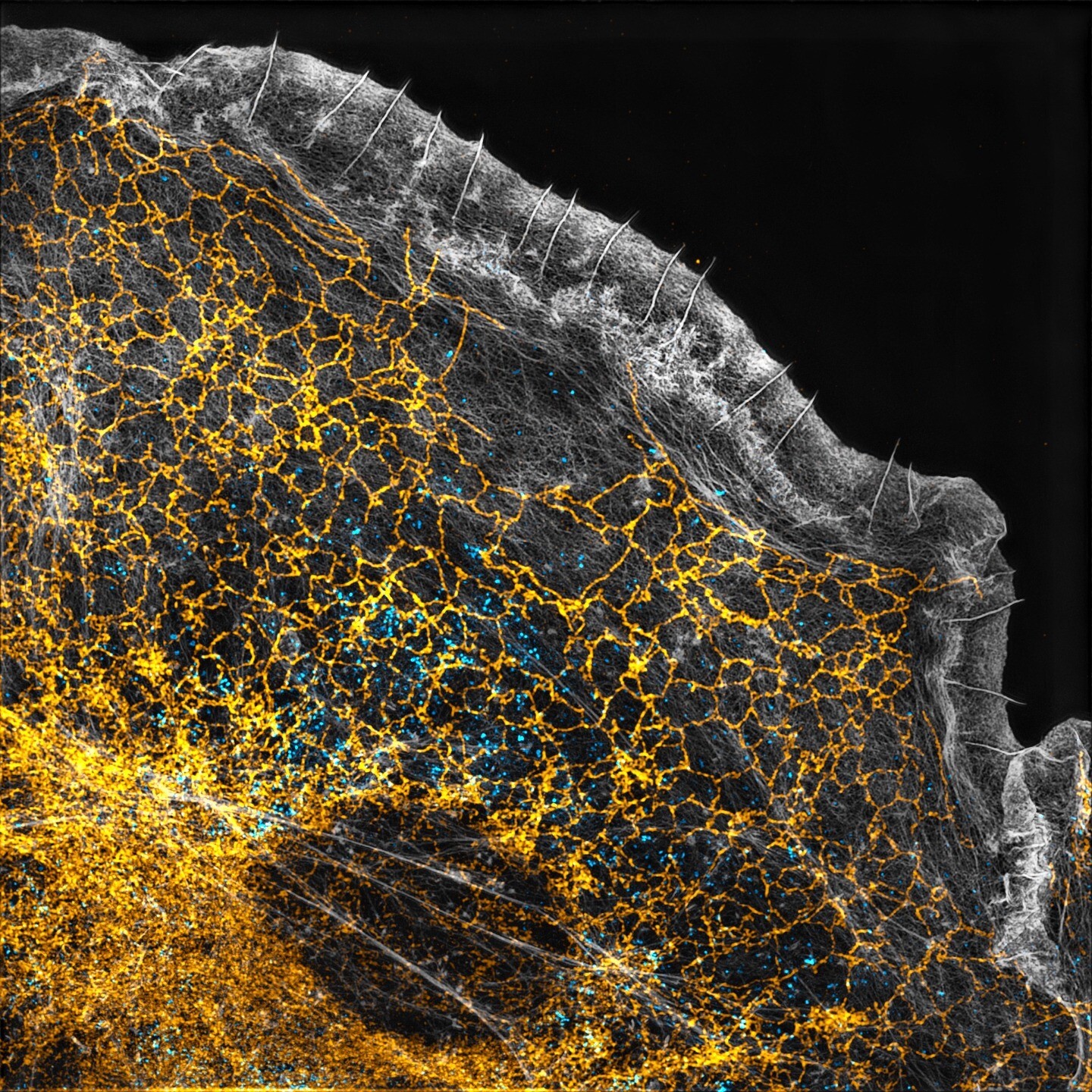
Moving toward a better #resolution: our structured illumination microscope (SIM) allows to get 2 times better detail than classical widefield or #confocal #microscopy. Here's a #cellfie of a monkey cell labeled for the endoplasmic reticulum (yellow), clathrin (blue) and #actin (gray) 🔬
#actin #cellfie #microscopy #confocal #resolution