
The EMBO Journal · @embojournal
555 followers · 290 posts · Server sciencemastodon.com
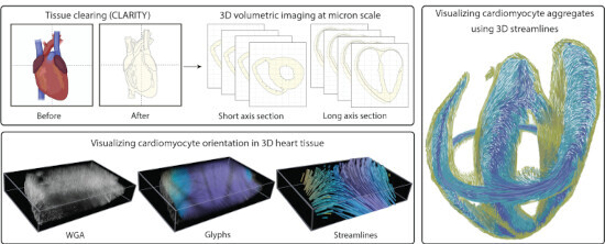
Cardiomyocyte orientation recovery at micrometer scale reveals long-axis fiber continuum in heart walls
A new method that combines #confocal #microscopy with computer vision provides unprecedented spatial resolution of #cardiomyocyte geometry along the mouse heart wall.
Minhajuddin Sirajuddin, Kaleem Siddiqi et al
#confocal #microscopy #cardiomyocyte

BioImagingUK · @BioImagingUK
41 followers · 216 posts · Server mstdn.science
RT @YorkBioimaging
4-Day, Hands-on Confocal Microscopy Course
https://www.york.ac.uk/biology/technology-facility/tfcourses/confocal-course/
Matured and evolved over 20 years and with 100% recommended delegate feedback.
Super fun, super interactive, and bonus being in the historic and amazing City of York.
#microscopy #imaging #confocal
#microscopy #imaging #confocal

Kay Schink · @kschink
141 followers · 208 posts · Server mstdn.scienceRT @YorkBioimaging
Last few places up for grabs. Inspiring, exciting samples, truely hands-on, great tutors and a wonderful city! Open to all.
Please retweet and pass on.
#microscopy #confocal https://twitter.com/YorkBioimaging/status/1618571443348082690

Open Microscopy Environment · @ome
82 followers · 10 posts · Server fosstodon.org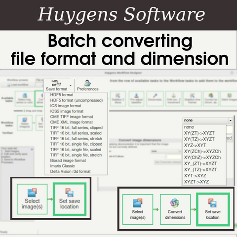
RT: @svi_huygens@twitter.com
#microscopists, want to get a better overview of microscopy image #FileFormats, bit depth, scaling, image #metadata, dimensions, and more?
Join our webinar Feb 23 and explore what formats e.g. #OMETIFF:
#microscopists #FileFormats #metadata #ometiff #microscopy #ImageAnalysis #confocal

Leica Microsystems · @leicamicrosystems
135 followers · 34 posts · Server mastodon.social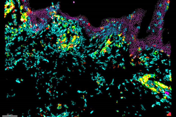
🔶 What are the additional insights you can gain from imaging tissue sample in 10 colors?
🔶 Follow our on-demand webinar with Dr. Nicolas Gaudenzio and discover how to perform 10-color imaging using a confocal microscope. See how this allows to assess the skin immune landscape.
#Sted #stellaris #confocal #immune

Biology Open · @BiologyOpen
309 followers · 55 posts · Server mstdn.science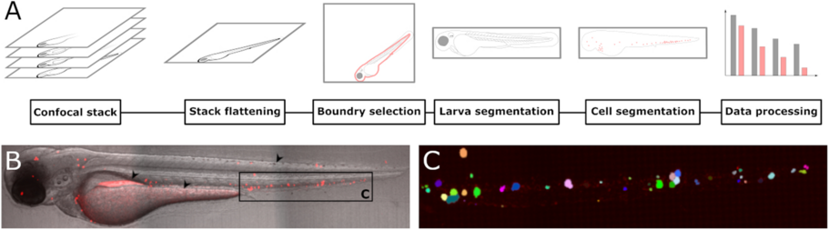
Jan-Lukas Førde et al. present a new #software tool for automatic #cell segmentation of fluorescent #cancer cells in #zebrafish larvae:
#Imaging #Biology #BiologyOpen #Science #AcademicMastodon #Confocal #Segmentation
#software #cell #cancer #zebrafish #imaging #biology #biologyopen #Science #AcademicMastodon #confocal #segmentation
Markus Osterhoff · @mosterh1
52 followers · 180 posts · Server academiccloud.socialTim Salditt (http://twitter.com/SaldittLab, substituting Jasper Frohn): #Multiscale #Xray Phase Contrast #Tomography at #GINIX/P10: Concepts, Implementation and Applications
- fantastic collaboration with the P10 team
- directly comparing #STED to #Minflux; old #confocal? not worth mentioning 😂
- overview tomo, then zoom-in
- tumorous human pancreatic tissue #biopsy: quantify the #tumor type
- from electron density to metrics, quantify fibres etc.
- #hippocampus patho punch, #Alzheimer sample
#alzheimer #hippocampus #tumor #biopsy #confocal #minflux #sted #ginix #tomography #xray #multiscale

Francesco Pasqualini · @fspasqualini
255 followers · 266 posts · Server fediscience.orgRT @Photometrics@twitter.com
Our thanks to Prof. @fspasqualini@twitter.com of @unipv@twitter.com who is using the #Kinetix #sCMOS, a @CrestOptics@twitter.com X-Light and a @NikonInst@twitter.com Ti2 for live #3D #spinningdisk #confocal #microscopy of #cardiac samples! Check out the full #Kinetix story here: https://www.photometrics.com/applications/customer-stories/pasqualini-live-cardiac-spinning-disk-pavia-kinetix
🐦🔗: https://twitter.com/Photometrics/status/1611384755144036354
#cardiac #microscopy #confocal #spinningdisk #3d #scmos #kinetix
Mike Schumacher PhD · @schumacher
127 followers · 18 posts · Server mstdn.science
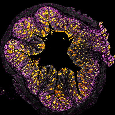
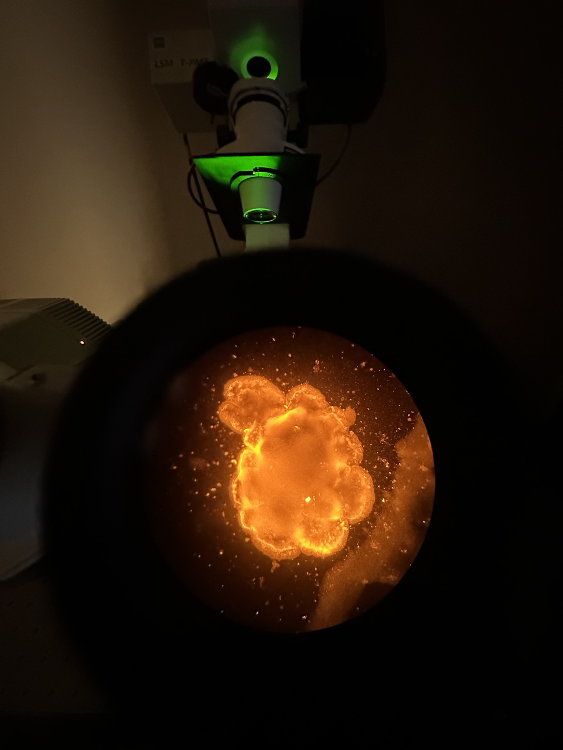
What’s better than looking down the #microscope at a new fiercely bright antibody that worked?
#organoids #sciart #immunofluorescence #confocal #microscopy #gi
#microscope #organoids #sciart #Immunofluorescence #confocal #microscopy #gi

Joseph P. · @tonic
132 followers · 277 posts · Server qoto.orgSkeletal #muscle is a type of tissue that makes up a large part of the human body. It is made up of many different cells that are able to contract and move. Skeletal muscle has the ability to #repair itself when it is damaged due to #aging, exercise, or diseases like #MuscularDystrophy. A small group of cells called #SatelliteCell s help with the repair process. Scientists have been trying to create models to study how #Skeletalmuscle develops and regenerates. Recently, they have been using human pluripotent #StemCell to create 3D models of skeletal muscle tissue. However, these models have not been able to recreate the full process of muscle regeneration. In this research paper, the authors introduce a new method of using human pluripotent stem cells to create 3D models of skeletal muscle tissue that can retain the ability to repair itself.
Over the past decades, scientists have used #animalmodel to study #muscleregeneration, which is regulated by #stemcell s. These animal models have been very helpful in understanding the mechanisms of muscle #regeneration, but they don't always accurately reflect the same range of diseases that humans experience. Therefore, researchers have suggested creating reliable in vitro models using human muscle cells. ( #hPSC s) could be used to create 3D human skeletal muscle #organoid s ( #hSkMO s) that contain sustainable #stemcell and distinct myofibers with the same proteins and structure as adult muscles. Previous approaches to skeletal muscle differentiation have been developed using 2D #culture systems, but these lack the natural environment and #StemCell niche that are necessary to model adult #myogenesis and muscle #regeneration.
#Stemcell s ( #SC s) can be used to repair damaged muscle tissue. They explain that SCs can be activated in response to muscle injuries and that other #cell types can contribute to the process of #myogenesis. The author then goes on to explain that #cytokine s, such as IL-4, can influence the #InflammatorySystem and promote SCs differentiation, which helps with muscle regeneration. While #organoid s generated from #hPSC s have potential, they do not fully replicate the in vivo native microenvironment. To address this, treat the #hSkMO s with extrinsic #cytokine s to promote #muscle #regeneration . #hSkMO s might then be used to study aspects of human muscle #biology and to identify novel #therapeutic candidates for muscle-wasting disorders.
To create a 3D structure of muscle tissue. They used #WNT activator and #BMP inhibitors at the beginning of the differentiation process to induce paraxial #mesodermal #cell s. They then added #FGF2 to the Matrigel to promote the 3D structure. #HGF and IGF1 were added later to accelerate the #myogenic specification and further #myofiber differentiation. They optimized the timing of the Matrigel embedding to day seven. After this, they observed #neuralcell s and withdrew FGF2 to focus on muscle tissue development. They then prolonged the HGF and IGF1 treatment to propagate #myogenic #progenitor s. They found that 62% of the #tissue was #skeletalmuscle tissue and that it contained PAX7+ #myogenic #stem / #progenitor cells, MYOD+ activated/committed #myoblast s, and MYOG+ #myocyte s. They also found that 31% of PAX7+/Ki67− and 29% of MYOD−/PAX7+ non-dividing quiescent SCs were present in the mature #hSkMO s. This indicates that the #hSkMO s were able to effectively recreate #embryo nic #myogenesis and have regenerative potential. Future studies using #singlecell #RNA sequencing may be necessary to further characterize the different types of cells in #hSkMO s.
The stepwise process to generate human skeletal muscle organoid s (hSkMOs) from human pluripotent stem cells (hPSCs)
The process begins with dissociating #hPSC s into #singlecell s and allowing them to form #embryoid bodies ( #EB s) in low-attachment V-shaped 96-well plates. Then, paraxial #mesodermal differentiation is promoted with #WNT activation, BMP inhibition, and FGF2 signaling. The expression of pluripotency markers OCT4 and NANOG decreases, and the expression of #mesoderm markers Brachyury, T-Box transcription factor 6 (TBX6), and mesogenin 1 (MSGN1) increases. To further characterize paraxial #mesoderm al differentiation, TBX6 is #immunostain ed. After paraxial #mesodermal induction, the #organoid s are embedded with growth factor-reduced Matrigel and transferred to a six-well plate on an orbital shaker. Growth factors are then added to the #myogenic specification media, and #hSkMO s are cultured until the day of analysis. The orbital shaker improves the viability, survival, and differentiation of hSkMOs by increasing the penetration rate of oxygen and nutrients into the core area of hSkMOs. The #hSkMOs gradually grow to more than 1.5 mm in diameter by day 60, appearing round-shaped, uniformly sized, and having relatively homogenous morphology. PAX3 and PAX7 are #myogenic progenitor markers, and their expression is verified by qRT-PCR and #cryo sections. The #myogenic cells appear as clusters, and approximately 9% of PAX7+ cells are double-positive for Ki67 at day 30, demonstrating that proliferating cells are #myogenic #progenitor s in hSkMOs. This indicates that the in vitro #hSkMO #culturesystem is able to recapitulate the features of embryonic skeletal #muscle development.
The different types of #SkeletalMuscle stem/progenitor cells that are involved in myogenesis, the process of muscle formation.
The researchers used qRT-PCR analysis and #immunohistochemistry to identify and characterize the different types of cells. They found that PAX3 and PAX7 (SC markers) were the major population during the early stage of #myogenesis, and that MYOD (proliferating and activated SC marker) and MYOG (differentiated myocyte marker) increased over time. They also observed that MYOD−/PAX7+, MYOD+/PAX7+, and MYOD+/Ki67+ cells accounted for 29%, 6%, and 8% of the putative quiescent, activated, and proliferating #SC s, respectively. MYOD+/PAX7− cells constituted 39% of differentiating myoblasts, and MYOG−/PAX7+ cells constituted 23% of putative quiescent SCs. MYOG+/PAX7− cells accounted for 30% of differentiated #myocyte s, and 8% and 6% of the MYOG+ cells in #hSkMO s co-expressed PAX7 and Ki67, respectively. This data shows that the researchers were able to identify and characterize different types of skeletal muscle stem/progenitor cells during #myogenesis.
The text is discussing the results of a research study that used hSkMOs (human skeletal muscle #organoid s) to study the development of skeletal muscle #tissue. The study found that the #hSkMO s grew exponentially in size within two months, and the growth rate then steadily decreased. The researchers then used scanning electron microscopy (SEM) imaging and confocal microscopy to examine the cytoarchitecture of the hSkMOs. They found that the hSkMOs contained a large population of terminally differentiated #myogenic cells and a small population of preserved myogenic stem/progenitor cells. They also found that the hSkMOs contained a substantial proportion of TITIN+ muscle cells and MAP2-positive #neuron s. To further characterize the presence of sustainable stem cells within the mature hSkMOs, they quantified the amount of dormant stem cells by #confocal #microscopy imaging. The results showed that approximately 56%, 31%, and 5% of PAX7+/Ki67- putative dormant stem cells existed throughout the differentiation of hSkMOs at days 30, 70, and 130, respectively. This indicates that the hSkMOs contained mature skeletal muscle properties and had the potential for #regeneration .
The researchers wanted to see if the #hSkMO s (human #skeletal muscle #organoid s) had the ability to regenerate #muscle #tissue after damage. To test this, they treated the hSkMOs with a cardiotoxin (CTX) which is known to induce muscle inflammation and damage. They then observed a decrease in PAX7+ and MYOD+ cells in the hSkMOs. To further test the #regenerative potential of the #hSkMO s, they added interleukin-4 (IL-4) to the medium to promote #muscleregeneration. After 14 days, they observed a significant increase in MYOG+ myocytes in the CTX-injured hSkMOs with the treatment of IL-4 compared to the CTX-injured hSkMOs without the treatment. This suggests that the hSkMOs have the potential to regenerate muscle tissue after damage.
Generation of Skeletal Muscle Organoids from Human Pluripotent Stem Cells to Model Myogenesis and Muscle Regeneration
Authors :
Min-Kyoung Shin , Jin Seok Bang , Jeoung Eun Lee , Hoang-Dai Tran , Genehong Park , Dong Ryul Lee and Junghyun Jo
#muscle #repair #aging #musculardystrophy #SatelliteCell #Skeletalmuscle #stemcell #AnimalModel #muscleregeneration #regeneration #hPSC #Organoid #hSkMO #culture #myogenesis #sc #InflammatorySystem #biology #therapeutic #Wnt #BMP #mesodermal #FGF2 #HGF #myogenic #myofiber #neuralcell #progenitor #tissue #stem #myoblast #Myocyte #embryo #singlecell #rna #embryoid #culturesystem #immunohistochemistry #neuron #confocal #microscopy #skeletal #regenerative #explainpaper #cell #cytokine #eb #mesoderm #immunostain #hSkMOs #cryo
Leica Microsystems · @leicamicrosystems
91 followers · 20 posts · Server mastodon.social
🔮 What insights could you gain from imaging tissues in 10-colors?
☑️ Join our upcoming webinar to discover how to perform 10-color acquisition using a confocal microscope and how this can help identify skin immune cells.
☑️ Register now https://fcld.ly/lbxrjmx
#confocal #immunecells #microscopy #ScienceTwitter #ScienceMastodon #science
#confocal #immunecells #microscopy #sciencetwitter #sciencemastodon #science

Alex_Kiryushkin · @Alex_Kiryushkin
45 followers · 7 posts · Server genomic.social
November lab work days :)
Here is a story continuation, what happened with cucumbers on Petri dishes from my previous tweet :)
Day one.
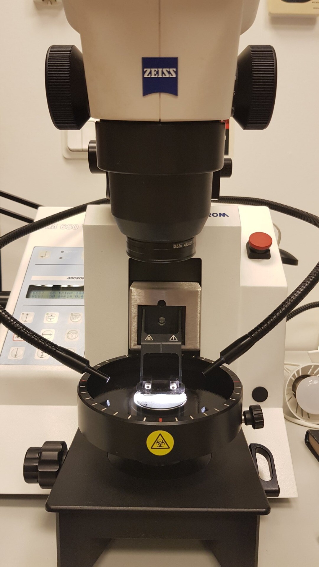
Preparing cross sections of cucumber transgenic root using vibratome.
#root #zeiss #cucumber
#vibratome
Day two.
Looking for:
1. native promoter expression in lateral root primordia (green nuclei) 2. auxin maxima (red nuclei) and stained cell walls (magenta).
Merge two pictures together :)
40×, single optical section, tile scan 2×2.
#root #LSM780 #confocal
#root #zeiss #cucumber #vibratome #lsm780 #confocal
Marta Perera · @MartaPerera
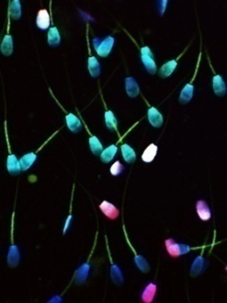
34 followers · 13 posts · Server mas.toThe future of #multispectral #imaging (and very nice images!) If you are acquiring only 4 #channels in the #confocal, welcome to the future! @Nature Congrats to the authors! https://www.nature.com/articles/s41596-022-00739-x
#confocal #channels #imaging #multispectral
Leica Microsystems · @leicamicrosystems
89 followers · 19 posts · Server mastodon.social
☑️ If you want to image more subcellular targets in parallel, this on-demand webinar “Live-cell fluorescence lifetime multiplexing using organic fluorophores” is right for you.
💹 You'll discover how you can expand your multiplexing capabilities using FLIM. Speaker is Dr. Michelle Frei.
https://fcld.ly/0kjk03c
#confocal #fluorescence #fluorophores #ScienceTwitter #ScienceMastodon
#confocal #fluorescence #fluorophores #sciencetwitter #sciencemastodon
Leica Microsystems · @leicamicrosystems
82 followers · 16 posts · Server mastodon.social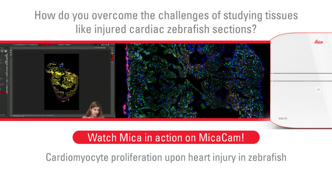
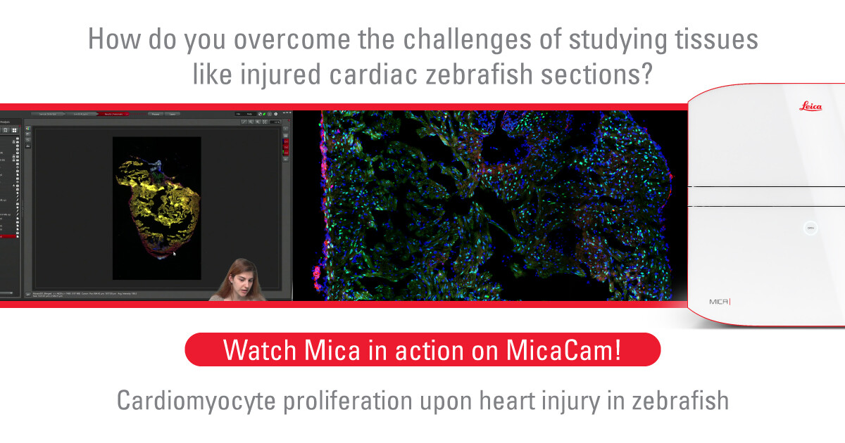
🖥💻 Learn about the biology of injured #cardiac tissue in #zebrafish. Find out how #Mica helps increase the efficiency of your workflow through easy navigation & exploration of the sections in #widefield followed by #confocal #imaging for a more detailed view.
https://fcld.ly/rgi74zp
#cardiac #zebrafish #mica #widefield #confocal #imaging #science #sciencetwitter #sciencemastodon #microscopy

Bruno C. Vellutini · @bruvellu
84 followers · 16 posts · Server mastodon.social
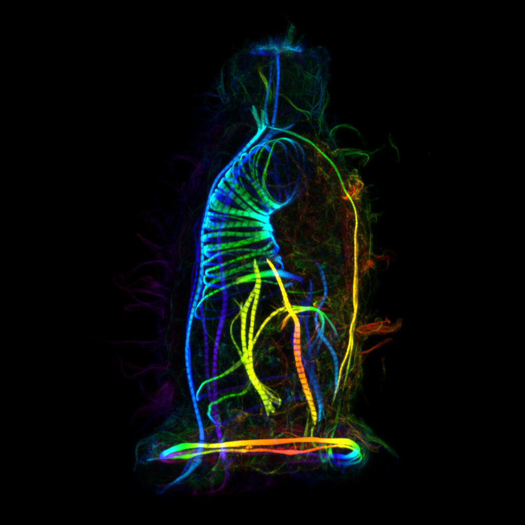
Today marks the 190th year anniversary of the discovery of the #cyphonautes #larva of #bryozoans! Ehrenberg first saw the tiny creature on November 25, 1832 (Hyman 1959).
This is my favorite #LarvalForm of all #MarineInvertebrates. It has a unique triangled, hat-like shape with a thin bivalve shell and a pompom at the top (apical organ). It swims and captures food using cilia and has some finely organized muscles (first image).
#cyphonautes #larva #bryozoans #larvalform #marineinvertebrates #otd #biology #bryozoa #microscopy #confocal #biodiversity

Andrew Burgess · @AndrewBurgessPhD
205 followers · 154 posts · Server mastodon.social
Throwback Thursday. #microscope #cellfie A mouse embryo fibroblast #MEF stained for #microtubule (blue) and #DNA (green). Images taken on a #Leica #confocal #SP8
#microscope #cellfie #mef #microtubule #dna #leica #confocal #sp8
Aurelie Dobric · @Aureliedobric
12 followers · 2 posts · Server mstdn.socialPs: I have a huge obsession for #confocal images and coffee 👍🏻
Now the introduction is fully complete !
Leica Microsystems · @leicamicrosystems
48 followers · 9 posts · Server mastodon.social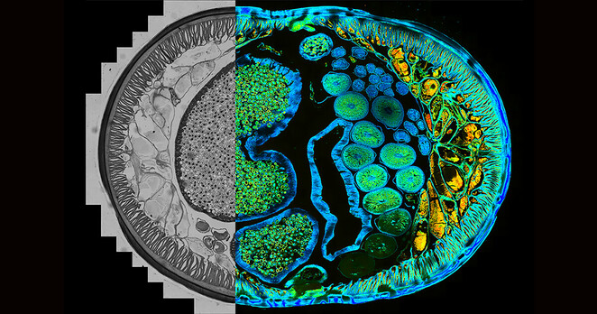
Understand how you can have a full sample overview with #confocal microscopy and quickly identify the important details instantly with the LAS X Navigator software.
👉 https://fcld.ly/egcph3k
📸🔬Identification of distinct structures in a transverse section of roundworm 🐛. The combination of the fluorescence lifetime-based information and the #fluorescence intensity with a stage application, gives an extra layer of contrast to tell different structures apart.
#confocal #fluorescence #sciencetwitter #sciencemastodon