
Elisabeth Kugler · @KuglerElisabeth
225 followers · 173 posts · Server mstdn.science
We love the point where science meets art.
What you see are cells of the eye that are important for neuron function and health. In the bottom half, where you see all the protrusions, is where the cells and neurons collide. #LoveScience
#lovescience #sciart #Science #ImageAnalysis #data #fluorescenceFriday

Moritz Negwer · @moritz_negwer
314 followers · 1733 posts · Server mstdn.science@lacouvee #FluorescenceFriday for pretty microscope pictures, if you're into that.
Also:
#fensterfreitag (german-speaking, but pictures of windows or views out of them should be pretty universal)
#caturday (cat pictures, just in case you need more of those)
There also is #TardigradeTuesday which doesn't have any content, but I wish it would. Hey #Tardigrade folks, this is your time to shine!
#fluorescenceFriday #Fensterfreitag #caturday #tardigradetuesday #tardigrade
Moritz Negwer · @moritz_negwer
294 followers · 1451 posts · Server mstdn.science@kagan may I suggest #FluorescenceFriday ? Lots of great microscopy pictures there
Elisabeth Kugler · @KuglerElisabeth
216 followers · 160 posts · Server mstdn.science
It's #FluorescenceFriday!
This week we look at the blood vessels in the brain of a 2-day-old #zebrafish using a state-of-the-art microscope. These data help scientists to understand how blood vessels form and develop.
#fluorescenceFriday #zebrafish #microscopy #BloodVessels #Science

Preston MacDougall · @ChemicalEyeGuy
283 followers · 8584 posts · Server mstdn.science“Chemical Eye 👁️ on Fool’s Amethyst” 👉 http://www.sitnews.us/MacDougall/092208_macdougall.html
#AmethystInitiative #DrinkingAge #BanFrats
#FluorescenceFriday
#amethystinitiative #drinkingage #banfrats #fluorescenceFriday

FocalPlane · @focalplane_jcs
362 followers · 181 posts · Server mstdn.science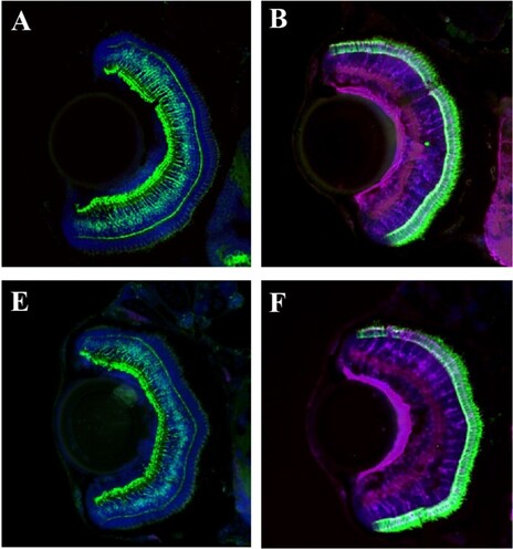
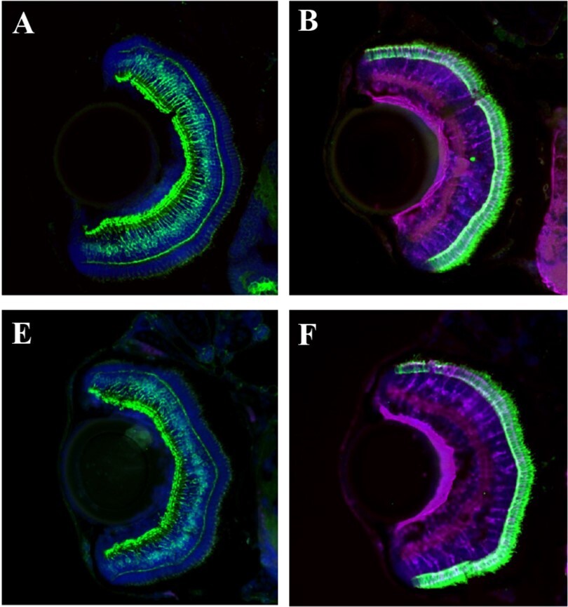
We’re celebrating #FluorescenceFriday by highlighting the #OpenAcess @BiologyOpen study from Siyu Yang, Mingzhe Cao and colleagues. They use #zebrafish to investigate retinal development and function and find that loss of flrt2 leads to microphthalmia.
#fluorescenceFriday #openacess #zebrafish
FocalPlane · @focalplane_jcs
361 followers · 168 posts · Server mstdn.science
Happy #FluorescenceFriday! Eunice Silva, Ana Venda and Catarina Homem find that serine hydroxymethyl transferase is required for optic lobe neuroepithelia development in Drosophila.
#fluorescenceFriday #devsimetabolism

Mary Fesenko · @MAFesenko
33 followers · 36 posts · Server mstdn.science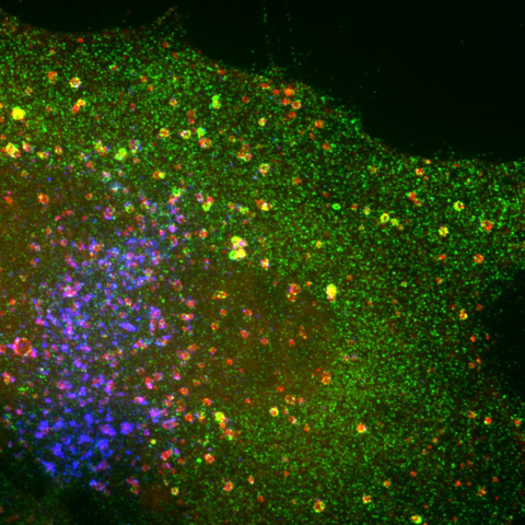
Happy #FluorescenceFriday! Imaging endosomes always brings me joy! 🔬🪩 #cellbiology #microscopy
#fluorescenceFriday #cellbiology #microscopy
FocalPlane · @focalplane_jcs
361 followers · 162 posts · Server mstdn.science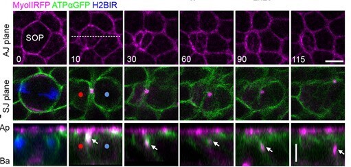
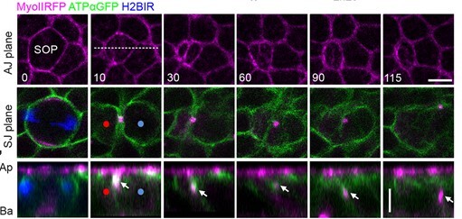
It’s #FluorescenceFriday and we’re looking at Drosophila SOP cytokinesis with Céline Bruelle, Roland Le Borgne and colleagues. They identify cell-intrinsic and -extrinsic functions of Shrub in SOP abscission.
📖@Dev_journal: https://journals.biologists.com/dev/article/150/10/dev201409/310743/Cell-intrinsic-and-extrinsic-roles-of-the-ESCRT
Moritz Negwer · @moritz_negwer
122 followers · 415 posts · Server mstdn.science
Hoang Anh Le (aka Anh), PhD · @AnhHLe2702
225 followers · 251 posts · Server mstdn.scienceHappy belated #FluorescenceFriday. Here is another video of a curious macrophage (in magenta) that went underneath an ectodermal cell (arrow) and split its nucleus in half to get through. The nucleus is also extremely plastic and it can deform significantly without rupturing!
Hoang Anh Le (aka Anh), PhD · @AnhHLe2702
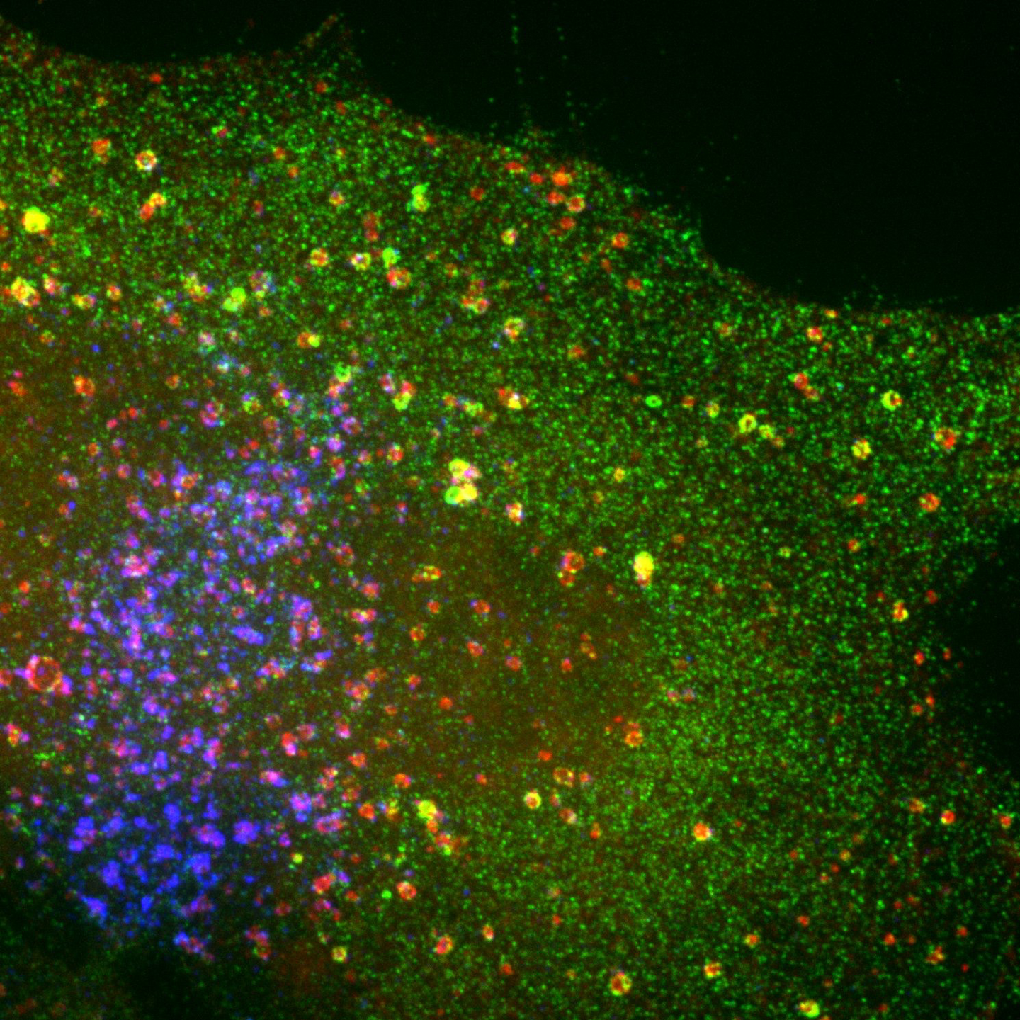
222 followers · 242 posts · Server mstdn.scienceI was looking through my old data from my PhD and came across this COS-7 cell with a rather interesting distribution of endosomes. They seem to localise just at the cell's lamella. Also, it is super bright. Happy #FluorescenceFriday!
FocalPlane · @focalplane_jcs
355 followers · 141 posts · Server mstdn.science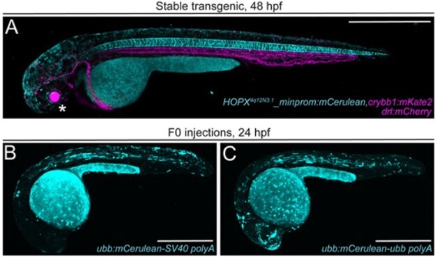
Hooray, it’s #FluorescenceFriday! This week we are admiring the beautiful work of Cassie Kemmler, @chrmosimann, Alexa Burger, Kristen Kwan and colleagues. They share their next-generation plasmids for transgenesis in zebrafish and more!
@Dev_journal: https://journals.biologists.com/dev/article/150/8/dev201531/307104/Next-generation-plasmids-for-transgenesis-in
Pablo J. Sáez :verified: · @pj_saez
431 followers · 130 posts · Server mstdn.scienceHere the result of the 1st test
---
RT @pj_saez
For a late #FluorescenceFriday
Expansion microscopy + SRFF = #ExSRFF
By @PuellesVictor lab and co.
https://www.nature.com/articles/s41565-023-01328-z #scicomm @focalplane_jcs https://twitter.com/pj_saez/status/1645885421367316482
https://twitter.com/pj_saez/status/1646990848419065861
#fluorescenceFriday #exsrff #scicomm
Pablo J. Sáez :verified: · @pj_saez
431 followers · 128 posts · Server mstdn.scienceFor a late #FluorescenceFriday
Expansion microscopy + SRFF = #ExSRFF
By @PuellesVictor lab and co.
https://www.nature.com/articles/s41565-023-01328-z #scicomm @focalplane_jcs
---
RT @pj_saez
#ExSRRF, a new pipeline that allows to reach 25 nm resolution using widefield #microscopy
(useful for #cellbio, #ER_literature, clinics & more)
Tks for the invitation, @PuellesVictor, happy that @cellcommlab we could help you w this neat work
#fluorescenceFriday #exsrff #scicomm #exsrrf #microscopy #cellbio #er_literature
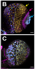
Hoang Anh Le (aka Anh), PhD · @AnhHLe2702
222 followers · 224 posts · Server mstdn.scienceThe other day, I noticed a few macrophages exploring their surrounding in a rather peculiar way. They press on the nucleus of the surrounding cells, hard enough to even deform and make it looks like they split the nucleus in half to get through. Check it out: #FluorescenceFriday

Jonas Wietek :vibing: · @JWietek
322 followers · 165 posts · Server mstdn.science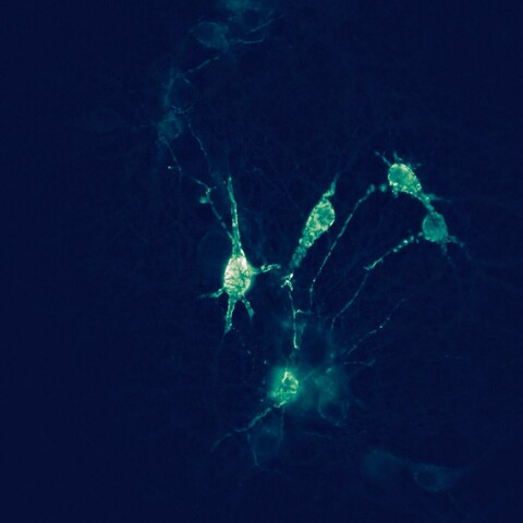
Hoang Anh Le (aka Anh), PhD · @AnhHLe2702
221 followers · 210 posts · Server mstdn.scienceRepost with a better gif: Can never get enough of these macrophages (in magenta) doing transcellular migration. Here is another example. You can also see a portion of the epithelial cell on top of its protrusion #FluorescenceFriday
Hoang Anh Le (aka Anh), PhD · @AnhHLe2702
221 followers · 207 posts · Server mstdn.scienceCan never get enough of these macrophages (in magenta) doing transcellular migration. Here is another example. It looks like the macrophage is probing at the cell-cell junction, but you can also see a portion of the epithelial cell on top of its protrusion #FluorescenceFriday
Hoang Anh Le (aka Anh), PhD · @AnhHLe2702
220 followers · 195 posts · Server mstdn.scienceRT @AnhHLe2702
Happy #FluorescenceFriday. As promised, here is a video of a macrophage (magenta) seemingly performing a transcellular migration through an ectodermal cell (cyan). It stops when it's half in and half out of the cell. Very blebby too. The event happens halfway through the video :)