Hoang Anh Le (aka Anh), PhD · @AnhHLe2702
220 followers · 197 posts · Server mstdn.scienceHappy #fluorescentfriday with another squeezy blebby macrophage (magenta) very eloquently manoeuvring through the complex architecture of the tissue (cyan). Look at those retraction fibres at its rear and the cytoplasm flow as it migrates. Master of morphodynamics.
Allan Carrillo-Baltodano · @allan_littlecar
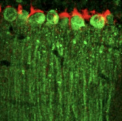
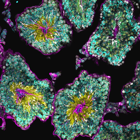
52 followers · 81 posts · Server ecoevo.socialSome antibodies arrived a few weeks ago, so we tested in Capitella. Looking forward to testing them in Owenia 🪱
Jonas Wietek :vibing: · @JWietek
320 followers · 154 posts · Server mstdn.scienceMeike van der Heijden · @meikeesther
101 followers · 8 posts · Server mastodon.worldFor this #FluorescentFriday, I want to share the most festive Purkinje cell of them all: Joy Zhou’s Purkinje cells with Santa hats.
Original image: https://elifesciences.org/articles/55569
Joy Franco · @engineeringjoy
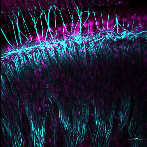
94 followers · 40 posts · Server mstdn.socialIt’s not #FluorescentFriday, but I spent my Saturday morning on the confocal so it’s worth posting about. Still tweaking immuno, imaging parameters, and processing, but so in love with the hearing organ that I have to stop and share.
#microscopylove #fluorescentfriday
Stacey Ogden 🦔 · @lab_ogden
245 followers · 93 posts · Server mstdn.scienceI love #fluorescentfriday ! This is an image of a developing mouse showing expression domains of one of our favorite genes, Sonic Hedgehog (SHH). SHH is a signaling protein that provides instructional cues that drive cell fate determination during developmental tissue patterning. Follow me for posts about cell biology, development, microscopy, lab antics, dogs, and an occasional yoga post. Happy Friday! 🦔
Fillip Port · @crisprflydesign
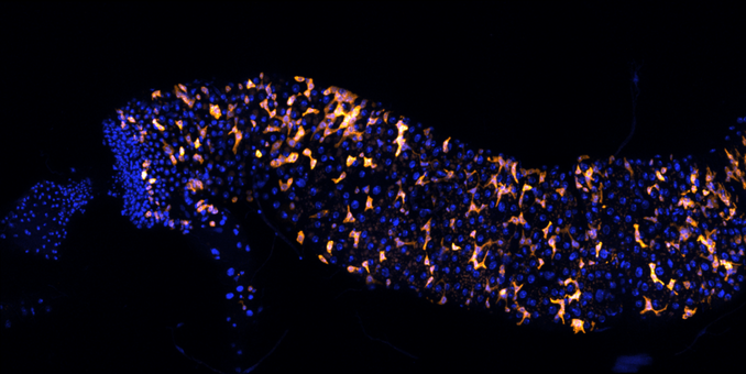
130 followers · 42 posts · Server fediscience.orgIts #flyday and #fluorescentfriday, so here is a Drosophila intestine with stem and progenitor cells in orange.
Mill_lab · @Mill_lab
301 followers · 165 posts · Server mstdn.socialHappy #FluorescentFriday folks! Because everything seems a little dark on the otherside, we are sharing some color-tastic eye-popping pics to kick off the week-end.... #cilia #flagella #MicroscopeMagic #TubulinCode
#tubulincode #microscopemagic #flagella #cilia #fluorescentfriday
Mill_lab · @Mill_lab
548 followers · 492 posts · Server mstdn.socialHappy #FluorescentFriday folks! Because everything seems a little dark on the otherside, we are sharing some color-tastic eye-popping pics to kick off the week-end.... #cilia #flagella #MicroscopeMagic #TubulinCode
#tubulincode #microscopemagic #flagella #cilia #fluorescentfriday
AIneurolab · @AIneurolab
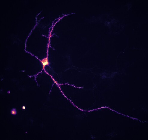
16 followers · 6 posts · Server mstdn.sciencePosting my first #fluorescentfriday pic on mstdn. Screen shot of cortical neurons genetically labeled with tdtomato.

