
Jonatan Hildén · @jhilden
443 followers · 2087 posts · Server vis.social
Fairly neat explorable 3D model of an ant’s mouth parts, built from a #microCT scan!
https://sketchfab.com/3d-models/leptomyrmex-uniclor-maxilla-muscles-8ca4b88c5c334ea68acb8089b8153097
By https://twitter.com/Formicadri/status/1693448003716350032

TU München · @tu_muenchen
357 followers · 240 posts · Server wisskomm.socialResearchers are using advanced #microCT to track the evolution of fungal cultures in #bioprocesses, revolutionizing our ability to optimize production and predict the growth of #fungi: https://go.tum.de/001896 🔬
#BiotechInnovation #Aspergillusniger
📷J.Hammel
#microct #bioprocesses #fungi #biotechinnovation #aspergillusniger

Dr. Nick Famoso · @DrFamoso
51 followers · 7 posts · Server ecoevo.social
@Boydpaleo MicroCT done... now to finish segmenting the little guy! #Hedgehog #paleontology #microCT #science
#hedgehog #paleontology #microct #science

Josh Gibson · @DrStrangeAnt
95 followers · 10 posts · Server toot.community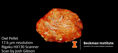
Josh Gibson · @DrStrangeAnt
95 followers · 9 posts · Server toot.community
Josh Gibson · @DrStrangeAnt
95 followers · 8 posts · Server toot.community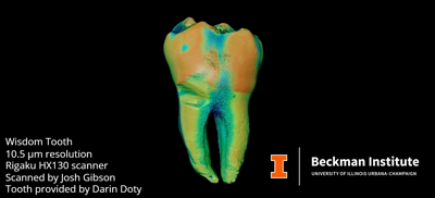
Fraunhofer IZFP · @Fraunhofer_IZFP
57 followers · 18 posts · Server wisskomm.social
Eine neue Peer-Review-Publikation mit Beteiligung unserer Wissenschaftlerin Sarah Fischer wurde erfolgreich veröffentlicht. Und das Gute daran: Sie ist open-access❗
A new peer-reviewed publication with the participation of our scientist Sarah Fischer has been successfully published. And the good thing about it: it's open-access❗
www.mdpi.com/2075-4701/13/2/201
#WeKnowHow #Fraunhofer #FraunhoferIZFP #metalmaterials #mechanicalengineering #adhesive #lasercutting #MicroCT @MDPI
#weknowhow #fraunhofer #fraunhoferizfp #metalmaterials #mechanicalengineering #adhesive #lasercutting #microct
April Neander · @aiNeander
119 followers · 64 posts · Server mastodon.artWork in progress! Version one of my next piece (I already know I’ll need to tweak it). Looks so cool all lit up undergoing UV curing! I just love how transmitted light works!
#wip #ExtraCTSkullpture #microct #ArtAndTech #3dprinting
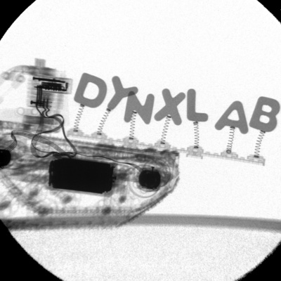
DynXLab · @dynxlab
9 followers · 3 posts · Server sciencemastodon.comWe have uploaded a short video to showcase our FleXCT facility here at the imec-Visionlab/University of Antwerp!
If you require any #microCT related services, do not hesitate to get in touch! https://uantwerpen.be/dynxlab

Daniel Pelliccia · @danielpelliccia
55 followers · 89 posts · Server aus.social
3D visualization of pores and inclusions by x-ray #microCT.
#microct #tomography #rocks #nondestructivetesting

IJPS · @IJPSJournal
117 followers · 26 posts · Server ecoevo.socialOur pleasure. It's a beautiful image.
---
RT @JJeiter
@IJPSJournal @VasileMarianna Thanks for choosing to put our image on the cover!
The 3D volume rendering on the cover as the rest of the #microCT data visualisation in the article was done with @OrsDragonfly3D .
https://twitter.com/JJeiter/status/1606780399946027008
Daniel Pelliccia · @danielpelliccia
50 followers · 76 posts · Server aus.social
Can you spot the problem with this LiPo #battery?
Here's a digital section of a battery obtained non-destructively with #microCT. Very easy to see the broken polymer layer in the middle.
#battery #microct #tomography #tomographytuesday
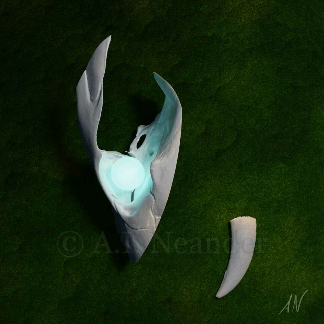
April Neander · @aiNeander
102 followers · 60 posts · Server mastodon.artAssembling the piece! I’m so excited! With all I’ve learned to make this piece a reality, I have so many ideas about where to go from here. Looking forward to making more art from #microCT data and exploring the beauty of #anatomy!
#3Dprinting #laserEngraving #smallElectronics #LEDArt #techArt #WIP #almostDone #ExtraCTSkullpture #sciart
#sciart #ExtraCTSkullpture #AlmostDone #wip #techart #ledart #smallelectronics #laserengraving #3dprinting #anatomy #microct

Vanderhoofius · @vanderhoofius
0 followers · 2 posts · Server masto.aiHi Mastodon!
I’m Brian Beatty (aka Vanderhoofius), a #paleontologist and #professor . I teach #anatomy and study #marinemammals . I’m rather fond of #surfacemetrology #histology and #microct . I particularly study #cetaceans #whales #sirenia #manatees #artiodactyla and #desmostylia (my favorite)!
#paleontologist #professor #anatomy #marinemammals #surfacemetrology #histology #microct #cetaceans #whales #sirenia #manatees #artiodactyla #desmostylia

DiceCT · @DiceCT
34 followers · 2 posts · Server ecoevo.socialIn this #OpenAccess pub, Kaylea describes a significant anastomotic network in the anterior spinal artery territory of the medulla –– we didn't know they were there, but Kaylea *found them* in super high-res #MicroCT scans of #DiceCT brains, and started wondering if maybe all that collateral (back-up) oxygen supply could explain why strokes of this artery are *super rare*

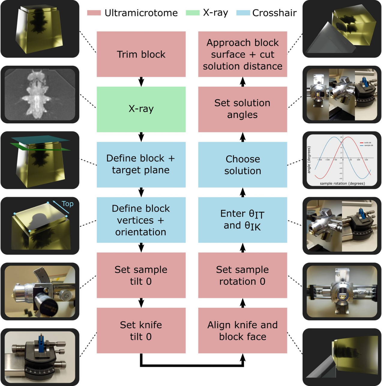
Albert Cardona · @albertcardona
728 followers · 626 posts · Server qoto.org
For all electron microscopists out there:
"Crosshair, semi-automated targeting for electron microscopy with a motorised ultramicrotome"
Kimberly Meechan et al. 2022 @eLife from Yannick Schwab's lab at EMBL in collaboration with The Crick institute. https://elifesciences.org/articles/80899
Presents a new method for reliably and "selectively targeting small regions of interest in a resin block by trimming with an ultramicrotome", powered by "user-friendly software to convert X-ray images of resin-embedded samples into angles and cutting depths for the ultramicrotome."
Reviewed by three outstanding electron microscopists: Christel Genaud, Song Pang, and Michaela Wilsch-Bräuninger.
#electronmicroscopy #microCT #Platynereis #science #methods #EMBL #TheCrick
#electronmicroscopy #microct #Platynereis #methods #embl #TheCrick #science
April Neander · @aiNeander
106 followers · 60 posts · Server mastodon.art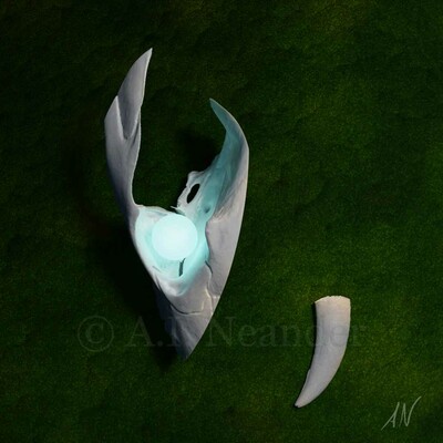
With a background in art and #microCT, it was only a matter of time before I combined the two. CT scans and the hidden anatomy within are beautiful, and ideas about what to do with those forms have been been bumping around in my head for a long time. After the pandemic started, I started working on what I call the ExtraCT Skullpture project, where I’m exploring different ways to use CT data to create digital and physical art. Not sure where this is going, but I'm having fun!

Phil Brailey-Jones (he/him) · @Pretzel
56 followers · 97 posts · Server ecoevo.social
RT @CarstenWMueller@twitter.com
PostDoc in my group in #Copenhagen – are you up to link #SoilStructure in the #Rhizosphere with the fate of #SoilOrganicMatter? We are looking for you to work with us from lab to field scale @science_ku@twitter.com @novonordiskfond@twitter.com #CarbonStorage #Imaging #MicroCT https://tinyurl.com/2tcx8kkh
🐦🔗: https://twitter.com/CarstenWMueller/status/1590646446767755264
#copenhagen #soilstructure #rhizosphere #soilorganicmatter #carbonstorage #imaging #microct
April Neander · @aiNeander
106 followers · 60 posts · Server mastodon.art
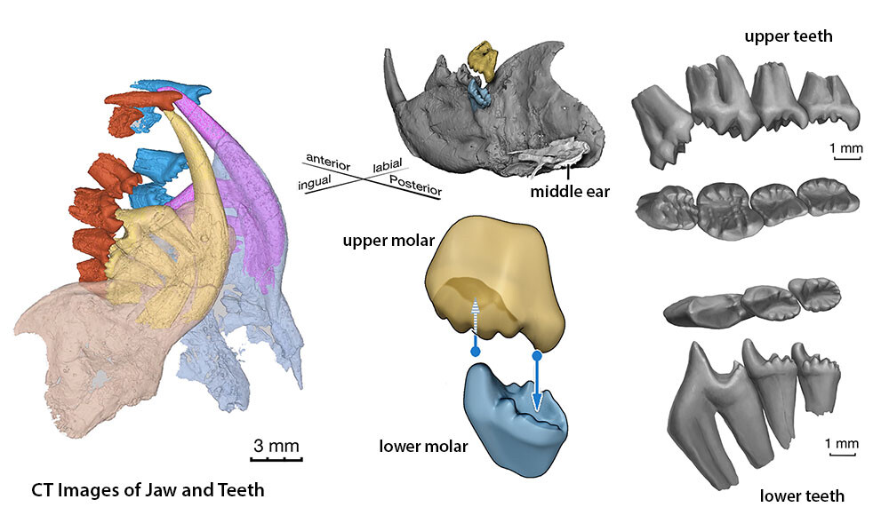
I also segment #microCT data for #3Dprinting and animation for the Luo Lab, where we study the transition from not-quite-a-mammal to true mammal. This means looking at a lot of teeth, ears and hyoids. Did you know the anatomical feature that defines a true mammal is three middle ear bones (incus, malleus, and stapes) that aren’t attached to the jaw!
April Neander · @aiNeander
106 followers · 60 posts · Server mastodon.artOur #microCT lab has contributed hundreds of scans to Morphosource via the oVert project. Researchers, educators, artists, etc. can download CT data from Morphosource free of charge! Its a great resource for anyone interested in #anatomy! https://www.morphosource.org/