
mau 🏳️🌈#PhaseOutFossilFuels · @mzedp
905 followers · 2770 posts · Server mas.toOne challenge of microphotography is that you can only get so much in focus at a time.
mau 🏳️🌈#PhaseOutFossilFuels · @mzedp
904 followers · 2767 posts · Server mas.to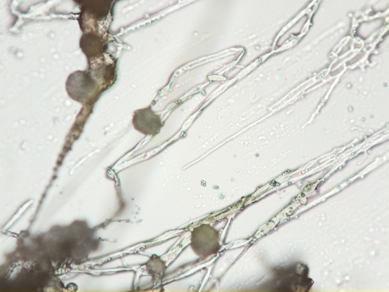
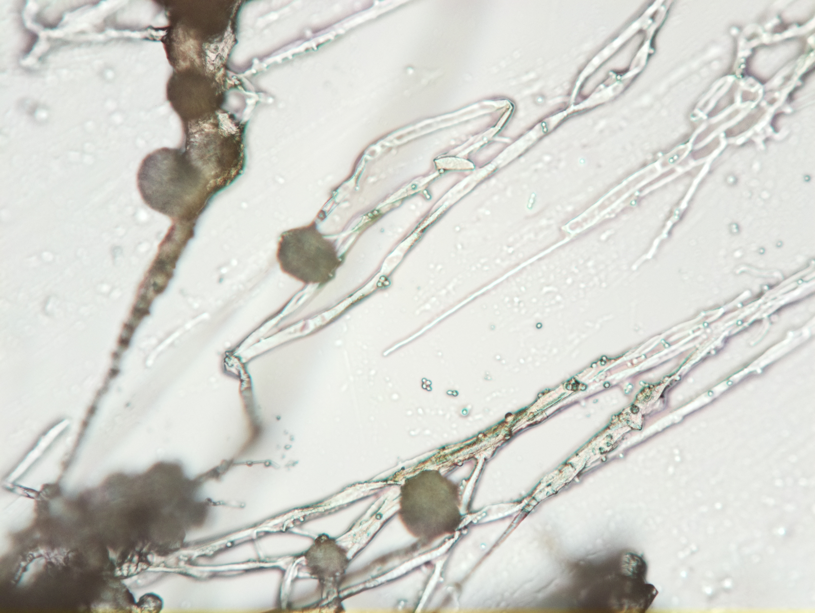
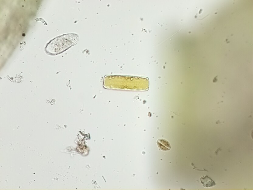
Bread got moldy
Normal person:*throws it out*
Weirdo with a microscope: "Huh, I wonder what that looks like up close"
I have no idea what this is, but I'll keep looking and try to figure it out.

📷 ᗰ⊙☋ᔕ†ꍏ☾♄ sun ⛅️ · @Moustachsun
90 followers · 518 posts · Server sunny.gardenLes “cascades de sang” en Antarctique : le mystère enfin résolu par la science
#Antarctic #mystery #microscopy

The EMBO Journal · @embojournal
555 followers · 290 posts · Server sciencemastodon.com
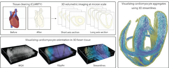
Cardiomyocyte orientation recovery at micrometer scale reveals long-axis fiber continuum in heart walls
A new method that combines #confocal #microscopy with computer vision provides unprecedented spatial resolution of #cardiomyocyte geometry along the mouse heart wall.
Minhajuddin Sirajuddin, Kaleem Siddiqi et al
#confocal #microscopy #cardiomyocyte

sumie-dh · @sumie_dh
13 followers · 11 posts · Server mstdn.science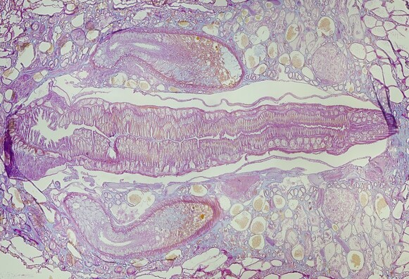
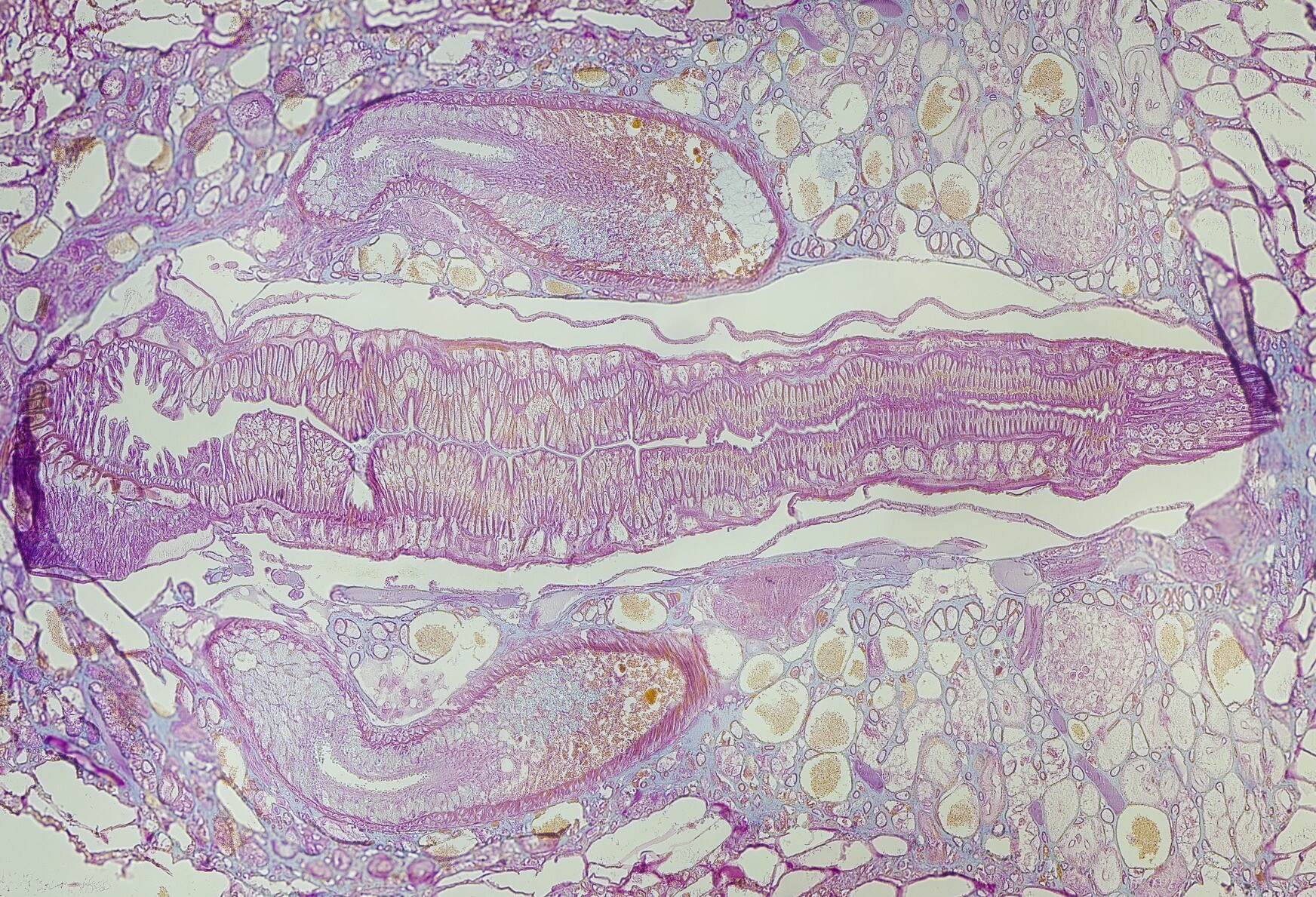
Glossiphonia sp #leech., frontal, Mallory–Heidenhain trichrome, 250x.That long noodle is proboscis - characteristic feature of Glossiphonidae. Note thin line around the proboscis - that's epidermis of proboscis poach.Two large croissant-like organs are ejaculatory ducts. Salivary glands are probably present on right side of the picture.Mystery: round organ which is present in pair at side of proboscis tip.Another mystery: yellow tissue inside of 'ducts'. #histology #sciart #microscopy #zoology
#leech #histology #sciart #microscopy #zoology
CellBioNews · @cellbionews
112 followers · 1836 posts · Server scientificnetwork.de
Researchers define protocol for high-resolution imaging of living #cells using atomic force #microscopy.
https://phys.org/news/2023-09-protocol-high-resolution-imaging-cells-atomic.html

philpem · @philpem
753 followers · 7973 posts · Server digipres.club
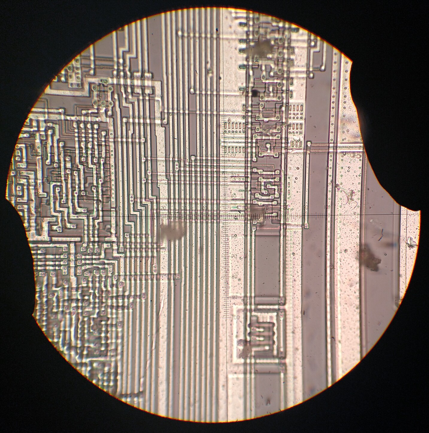
Comparison of Brightfield versus DIC illumination, Motorola 68HC05 microprocessor.
The quality here is fairly poor; my light source polariser is a piece of cheap polarising film clipped into a 3D-printed frame.
I need to find somewhere that sells good quality ~18 to ~22mm glass polarizers.
#ICRE #microscopy #reverseengineering
#reverseengineering #icre #microscopy

Human Technopole · @humantechnopole
341 followers · 112 posts · Server mstdn.science
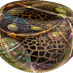
🔬 How to unveil a Chlamy organisation without expensive instruments and specific training?
Our Pigino Group with the University of Geneva, developed an ExM-based protocol to visualise the 3D ultrastructural organisation of Chlamydomonas reinhardtii by using conventional optical light microscopes.
✌️ Published in Bio-protocol and available now to the research community.
#lifesciences #humantechnopole #microscopy #science #sciencemastodon
#lifesciences #humantechnopole #microscopy #Science #sciencemastodon
Ignacio Izeddin · @IzeddinResearch
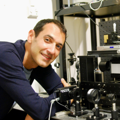
12 followers · 7 posts · Server fediscience.orgAre there any microscopists hiding here?
Adding :AddBiophysicist to this post in order to get in the "Biophysicists on Mastodon" list...

Andrew Plested · @andrewplested
293 followers · 1231 posts · Server mstdn.science
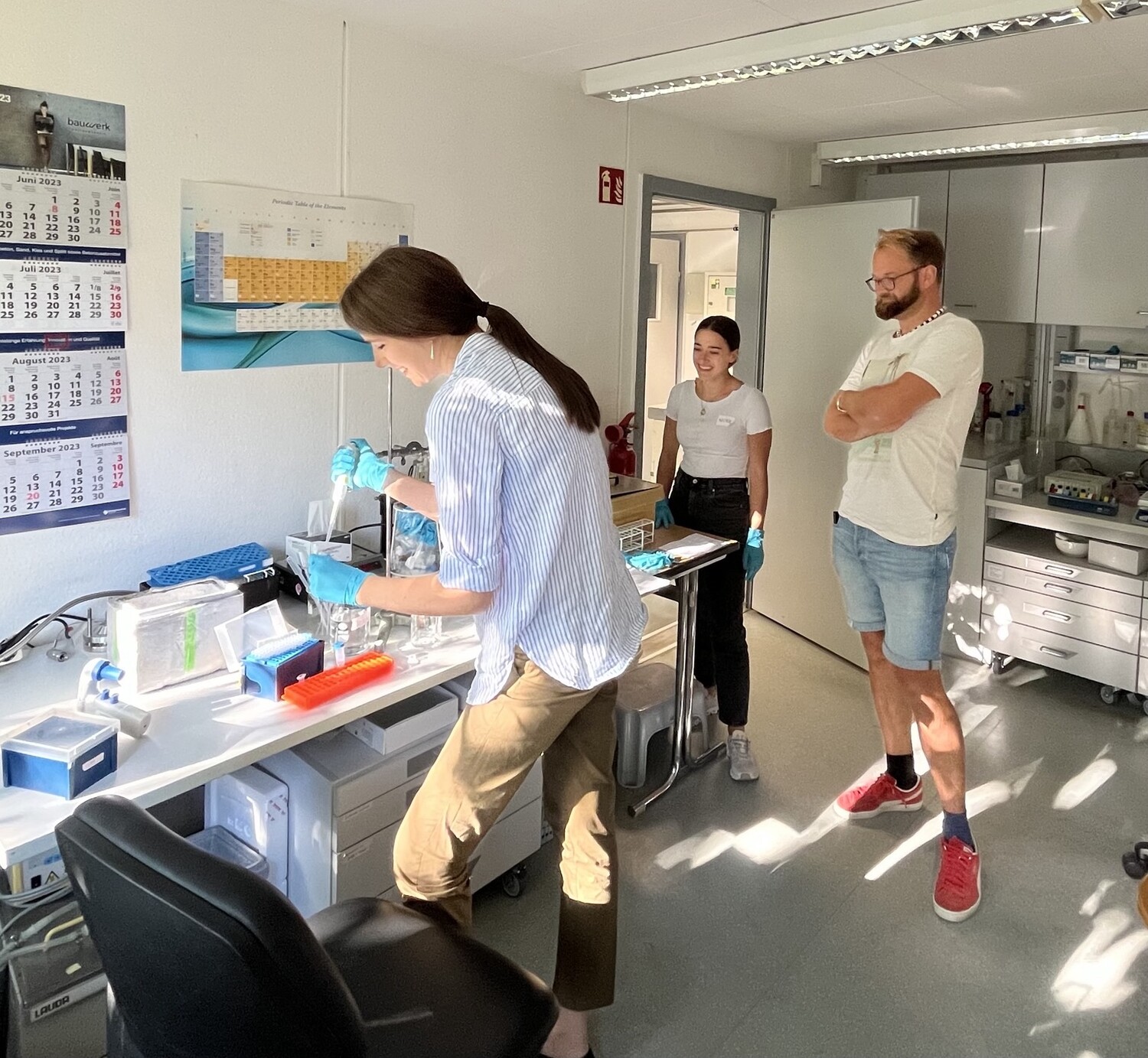
Iva Lučić shows our students how to label CaMKII with fluorescent dye for #singleMolecule TIRF #microscopy #biophysics
#singlemolecule #microscopy #biophysics
mau 🏳️🌈#PhaseOutFossilFuels · @mzedp
856 followers · 2658 posts · Server mas.toNice, finally got the video recording option working so now I can also try my hand at microvideography.
#openflexure #videography #microscopy #biology

Bose-Einstein-Kondensat · @MWNautilus
111 followers · 16 posts · Server mstdn.social
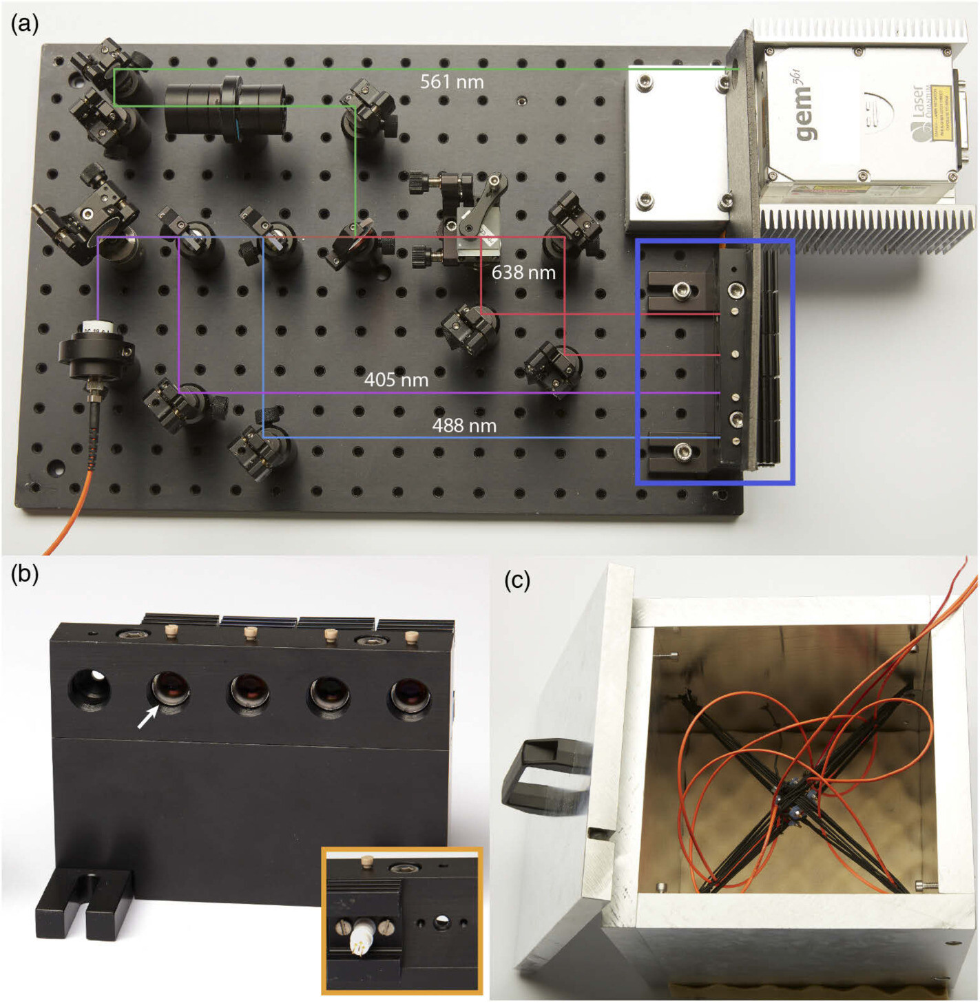
Cost-efficient #OpenSource #laser engine for #microscopy:
-405 nm, 488 nm & 638 nm and optionally a 561 nm diode-pumped solid-state laser
-agitated multimode fiber to suppress speckles
https://doi.org/10.1364/boe.380815
#DIYbio #hackerspace #lab #instruments #SMLM #microscope #imaging
#imaging #microscope #smlm #instruments #lab #hackerspace #diybio #microscopy #laser #OpenSource
Bose-Einstein-Kondensat · @MWNautilus
111 followers · 16 posts · Server mstdn.social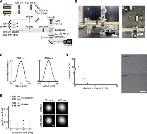
Simple, economic & robust rail-based #OpenSource setup for multicolor super-resolution localization #microscopy:
https://pubs.acs.org/doi/10.1021/acs.jpca.3c01351
#DIYbio #lab #instruments #microscope #SMLM #imaging
#imaging #smlm #microscope #instruments #lab #diybio #microscopy #OpenSource

Moritz Negwer · @moritz_negwer
314 followers · 1733 posts · Server mstdn.science@PhiloNeuroScie Nice find! #Tissueclearing and #lightsheet #microscopy work on human brain (sections) too :)
Human brain samples are normally quite hard to properly image because they are dense with lipofuscins, super autofluorescent and not very permeable to antibodies. That they managed to pull this off using the more-or-less standard iDISCO protocol (even with "just" a 4 mm thick slab and high antibody concentrations) is quite impressive.
edits: corrected thickness + typos
#tissueclearing #lightsheet #microscopy

Leibniz-HKI · @LeibnizHKI
49 followers · 34 posts · Server wisskomm.social
Tomorrow, the Rockin' Science Tour by Miltenyi Biotec will make a stop at the Leibniz-HKI 🚛
From 11 a.m. to 4 p.m, talk to experts about cell culture🧫, flow cytometry and more🦠🔬!
Or just stop by during your lunch break to play a few games and win some prizes🎈
#ScienceFestival #Science #microbiology #microscopy #cellbiology #cellculture
#sciencefestival #science #microbiology #microscopy #cellbiology #cellculture
sumie-dh · @sumie_dh
13 followers · 9 posts · Server mstdn.science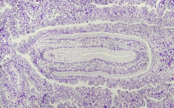
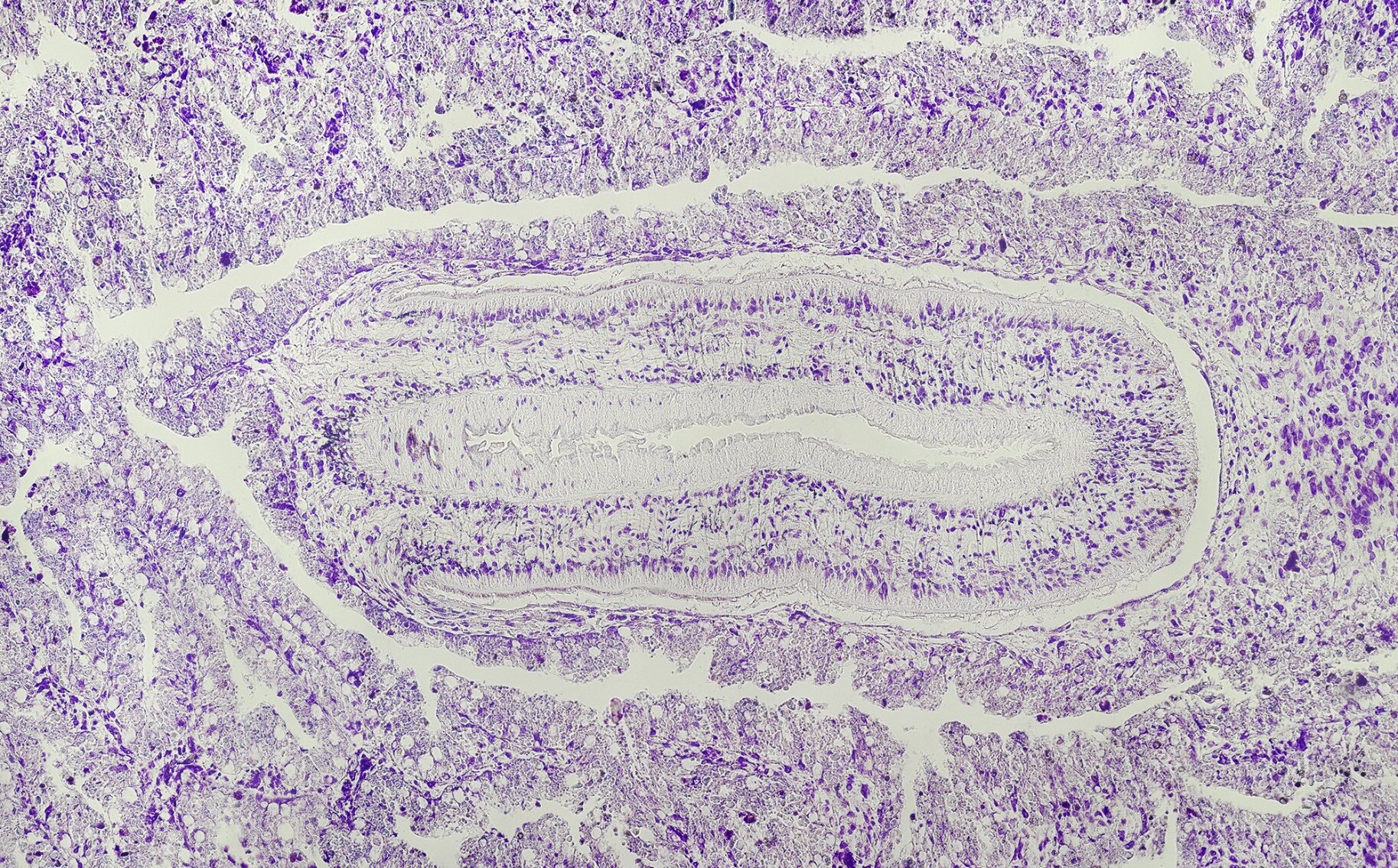
Byproduct of brain hunt [G. tigrina x G. sinensis morphology examination] - pharynx of Girardia sp., cresyl acetate, frontal - 8 frames, 250x. All 3 branches of intestine are visible as well -2 are visible as long lumens alongside of the pharyngeal pouch. #histology #planaria #planarian #microscopy #zoology
#histology #planaria #planarian #microscopy #zoology
sumie-dh · @sumie_dh
11 followers · 7 posts · Server mstdn.science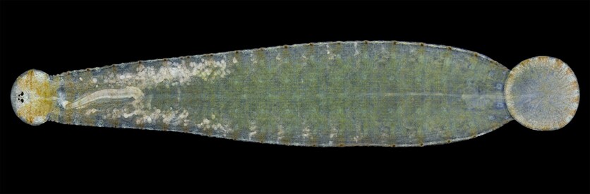
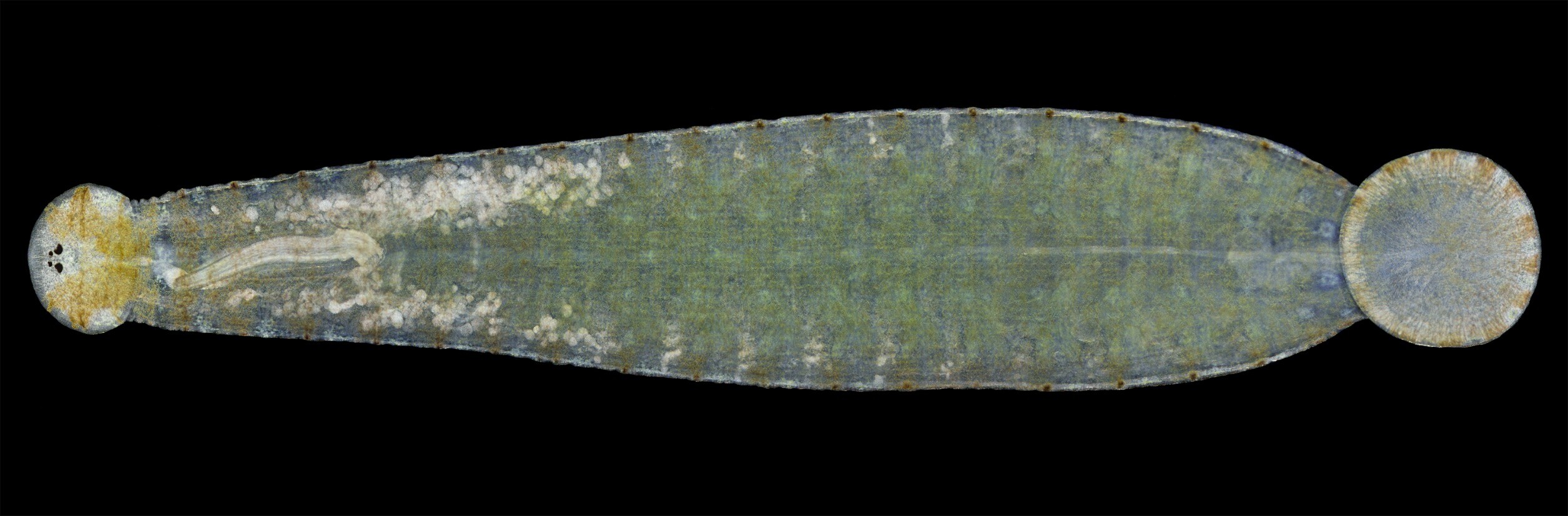
Hemiclepsis marginata - #leech , whole mount, cca 20 frames, 100x, pseudo-dark field
#leech #microscopy #meiofauna #zoology
mau 🏳️🌈#PhaseOutFossilFuels · @mzedp
847 followers · 2607 posts · Server mas.to
What good is a microscope in the hands of an amateur?
Well, it makes a drop of dirty water infinitely more interesting than it would be otherwise.
Now I've just gotta learn to identify what these things are.

Elisabeth Kugler · @KuglerElisabeth
218 followers · 166 posts · Server mstdn.science
Did you know?
Scientists can study how cells move, form, and interact in real-time using state-of-the-art microscopes.
In this picture you see cells of the eye developing from the outside to toward the lense. Studying their movement helps scientists understand cell interactions, development, and health.
Elisabeth Kugler · @KuglerElisabeth
218 followers · 166 posts · Server mstdn.science
Business values provide the compass for our journey, ensuring unwavering (1) quality, (2) respect, (3) courage, (4) integrity, and (5) focus. They create the foundation for trust, growth, and a purpose-driven path to success.
We will have some more on this tomorrow. Watch out for more!
#business #businessvalues #smallbusiness #businessgrowth #success #quality #respect #courage #integrity #focus #microscopy #science #art #sciArt
#business #businessvalues #smallbusiness #businessgrowth #success #quality #respect #Courage #integrity #focus #microscopy #Science #art #sciart