Furqan Shah · @furqanshah
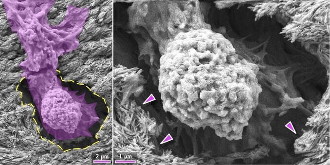
51 followers · 85 posts · Server mstdn.scienceAlien invasion!!! 👾 👽 Under in vitro culture, a #human #mesenchymal #stemcell crawls into an #osteocyte lacuna (http://bit.ly/Osteocyte) at the #bone surface, observed by scanning #electron #microscopy 🔬 🦴 Forget shape-memory #alloys! #Cells remember! (Arrowheads = bone #mineral)
#biology #electronmicroscopy #stemcells #imaging #bioimaging #biomimicry #biomineralization #invitro #apatite
#human #mesenchymal #stemcell #osteocyte #bone #electron #microscopy #alloys #cells #mineral #biology #ElectronMicroscopy #stemcells #imaging #bioimaging #biomimicry #biomineralization #invitro #apatite
Furqan Shah · @furqanshah
51 followers · 82 posts · Server mstdn.scienceImaging #bone with scanning electron microscopy? 🔬
Correlate – know better! 🦴👀
The combination of backscattered electron (BSE) and secondary electron (SE) imaging modes reveals a more accurate view of bone around #osteocyte lacunae, the small spaces where bone cells reside. 🔍💀 This information is crucial for understanding bone remodelling and how bone responds to injury or disease. 🤔📈
Read more:
https://nature.com/articles/s41413-019-0053-z
#biomaterials #electronmicroscopy #science #biology #physics #bioimaging
#bone #osteocyte #biomaterials #ElectronMicroscopy #Science #biology #physics #bioimaging
Furqan Shah · @furqanshah
51 followers · 78 posts · Server mstdn.science#Magnesium #whitlockite formation in the alveolar #bone, with #bisphosphonate exposure and osteolytic #skeletal #metastasis. Scanning electron microscopy, Micro-Raman spectroscopy, and energy dispersive X-ray spectroscopy reveal Mg-rich, rhomboidal nodules (200 nm to 2.4 µm) within the lacuno-canalicular space. Mg-whitlockite formation in #osteocyte lacunae is multifactorial in #nature and suggests altered bone #biomineralization.
#magnesium #whitlockite #bone #bisphosphonate #skeletal #metastasis #osteocyte #nature #biomineralization #biology #pathology #Science
Furqan Shah · @furqanshah
45 followers · 73 posts · Server mstdn.scienceThrough the eyes of an #osteocyte. In #bone, this is what the environment of partially embedded osteocytes (osteoblastic–osteocytes) is like!
Scanning electron microscopy has been used to observe the orientation of mineral platelets in relation to osteoblastic–osteocyte lacunae on the surface of deproteinized trabecular bone in adult sheep.
Read more:
https://doi.org/10.1007/s00223-015-0072-8
@zeiss_micro #microscopy #biology #imaging #biomineralization #extracellularmatrix #friday
#osteocyte #bone #microscopy #biology #imaging #biomineralization #extracellularmatrix #friday
Furqan Shah · @furqanshah
45 followers · 73 posts · Server mstdn.science#Magnesium #whitlockite formation in the alveolar #bone, with #bisphosphonate exposure and #osteolytic #skeletal #metastasis. Scanning electron microscopy, Micro-Raman spectroscopy, and energy dispersive X-ray spectroscopy revealed Mg-rich, rhomboidal nodules (~200 nm to ~2.4 µm) within the lacuno-canalicular space. Mg-whitlockite formation within #osteocyte lacunae is multifactorial in #nature and suggests altered bone #biomineralization.
#magnesium #whitlockite #bone #bisphosphonate #osteolytic #skeletal #metastasis #osteocyte #nature #biomineralization #biology #pathology
Furqan Shah · @furqanshah
45 followers · 73 posts · Server mstdn.scienceHere's a transmission electron microscopy (TEM) image of #bone showing mineralized #collagen and an #osteocyte lacuna containing microcalcifications of #apatite and #whitlockite.
Such #calcification or #mineral formation is part of normal physiological processes such as #apoptosis but can also indicate #pathology.
The specimen was prepared using focussed ion beam scanning electron microscopy (FIB-SEM).
Read more:
https://pubs.acs.org/doi/abs/10.1021/acs.nanolett.7b02888
#bone #collagen #osteocyte #apatite #whitlockite #calcification #mineral #apoptosis #pathology #ThinSectionThursday #biomaterials #ElectronMicroscopy


