
Petra Kaminen Mosher · @kaminenmosher
425 followers · 1389 posts · Server zirk.us
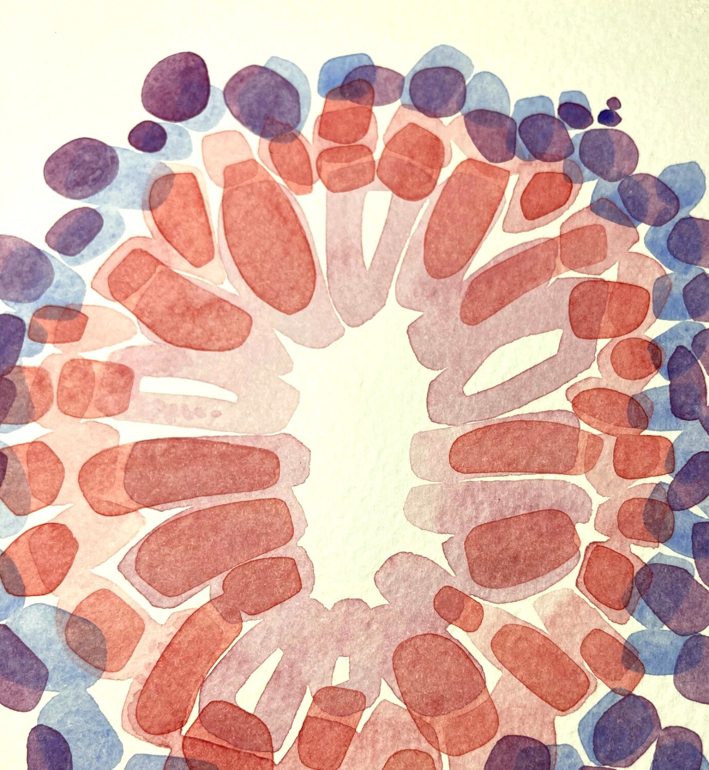
Gastric gland cells from a #pathology slide as #WatercolourPaintings from 2017. Available at my website shop https://kaminenmosher.com/direct-from-the-studio
#painting #art #PathArt #ContemporaryPainting #microscopy #watercolour #watercolor #InspiredByScience #SciArt
#pathology #watercolourpaintings #painting #art #pathart #contemporarypainting #microscopy #watercolour #watercolor #inspiredbyscience #sciart

Shane Battye · @shanebattye
195 followers · 307 posts · Server aus.social
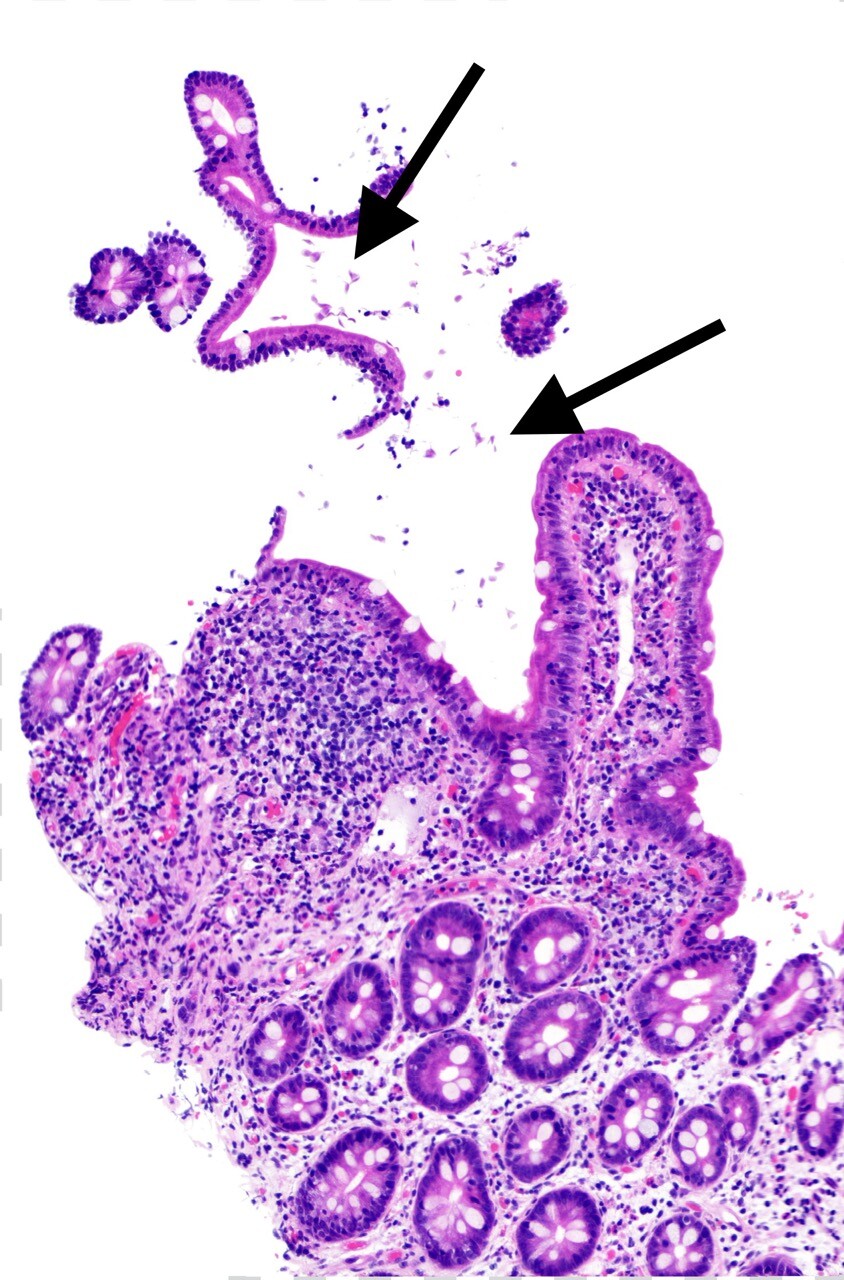
Here’s a case of duodenal #Giardiasis (causative organism Giardia lamblia) at 200x magnification. The #Giardia organisms are somewhat tear-drop shaped and resemble falling leaves, overlying the surface of the duodenum (the first segment of small bowel after the stomach). The duodenal mucosa is inflamed.
#pathology #pathart #bstpath #pathmastodon #GIpathology #InfectiousDisease
#pathology #pathart #bstpath #pathmastodon #gipathology #infectiousdisease #giardiasis #giardia

Shane Battye · @shanebattye
128 followers · 167 posts · Server aus.social@paravatar a few on med-mastodon.com server and try #pathology #bstpath #pathart #medmastodon #pathmastodon
#pathology #bstpath #pathart #MedMastodon #pathmastodon
Shane Battye · @shanebattye
128 followers · 167 posts · Server aus.social
You’ve heard of benign fibrous histiocytomas and their cutaneous (skin) equivalent aka dermatofibroma or “DF” for short (very common small skin lesions predominately on arms/legs)
BUT have you heard of an #EpithelioidFibrousHistiocytoma ? These are still benign but can look a bit scary under the microscope - big cells, big nuclei, big nucleoli, multinucleated cells, few mitoses for good measure 😐
Here’s one under my #microscope at 100x (just showing the top of the lesion as I didn’t have time to scan the whole thing for this toot - the area under the black line in the second pic), the dense pink band at the top is the epidermis - the part of skin you can touch).
Not shown are the immunostains I also used (positive for F13a, some scattered CD68 cells; negative for SOX-10 (melanoma marker) and pancytokeratin (carcinoma marker); vascular stains only highlighted vessels). And I showed it to a colleague too, to ensure we’re in agreement.
#epithelioidfibroushistiocytoma #microscope #pathology #bstpath #pathart #MedMastodon #pathmastodon
Shane Battye · @shanebattye
128 followers · 167 posts · Server aus.social
A really florid case of #endometriosis down my #microscope on low power magnification. This is a cross section of small bowel (the compressed lumen visible on the right). The endometrial glands (left of my black line) are expanding and distorting the muscular wall of this segment of bowel. There are fibrovascular adhesions overlying the outside surface (the serosa) of the bowel.
Endometriosis is the presence of endometrial tissue outside the uterus (most often around the ovaries and adjacent structures - but can occur anywhere). It causes significant morbidity from pain, infertility and an associated risk of ovarian cancer.
#endometriosis #microscope #pathology #bstpath #pathart #MedMastodon #pathmastodon