
Furqan Shah · @furqanshah
68 followers · 116 posts · Server mstdn.science
🔬🏖️ Grains of #sand under a scanning electron #microscope – varied shapes, textures, vibrant microstructures. In the backscattered electron (BSE) #imaging mode, bright spots indicate presence of higher atomic number elements or higher average atomic density 🔍 #Microscopy #MicroscopicWorld #ElectronMicroscopy #ScanningElectronMicroscopy #Small
#sand #microscope #imaging #microscopy #microscopicworld #ElectronMicroscopy #scanningelectronmicroscopy #small
Paul Harnik · @paulharnik
219 followers · 25 posts · Server ecoevo.social
Exploring #TinyWorlds today with our SEM. Here are Nucula proxima & Nuculana acuta, 2 marine bivalves that live in the N. Gulf of Mexico; Top left is N. acuta, top right is N. proxima, lower left is a close-up of N. proxima, and lower right of N. acuta. Close-ups show their lovely larval shells! #ScanningElectronMicroscopy
#tinyworlds #scanningelectronmicroscopy
Furqan Shah · @furqanshah
45 followers · 73 posts · Server mstdn.science
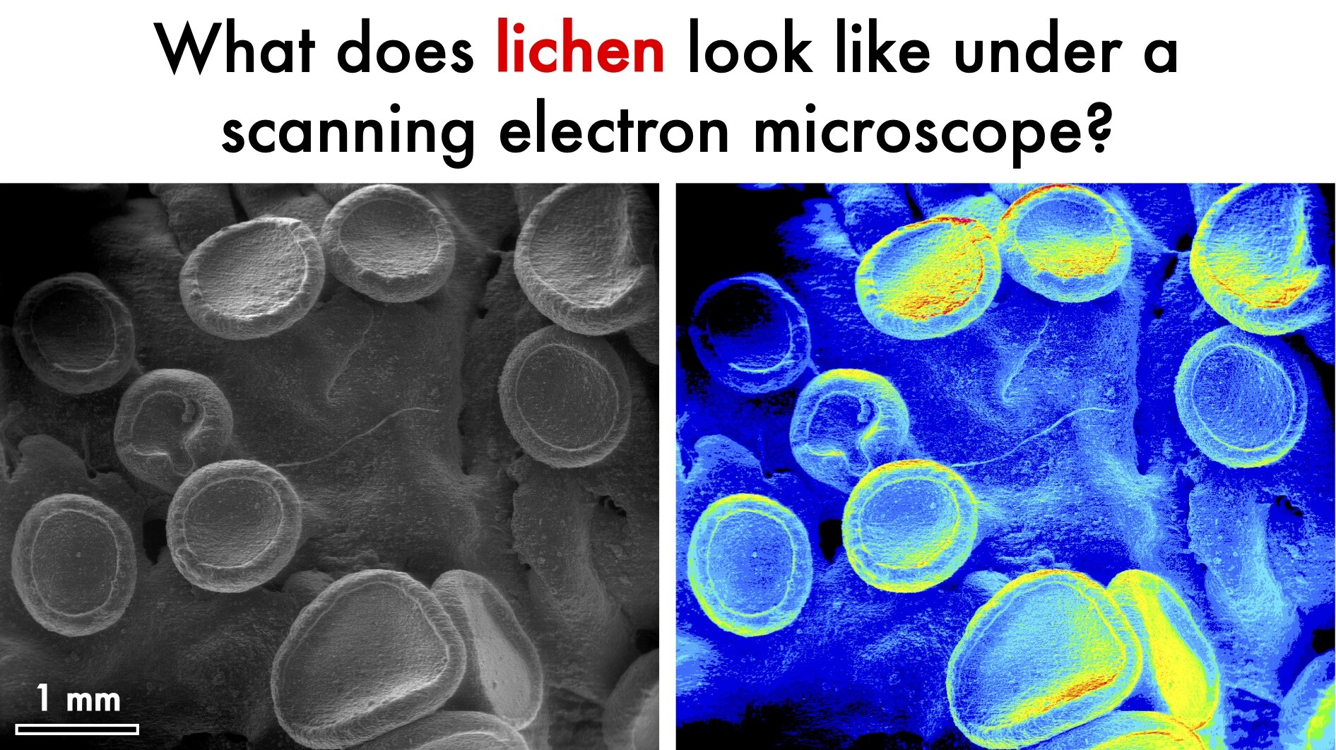
What does #lichen look like under a scanning electron microscope? 🔬
Look no further! 🌲🌿🌱🦠🍀
#nature #moss #algae #bacteria #fungi #cyanobacteria #trees #botany #ecology #biodiversity #trees #biology #botany #winter #snow #photo #photooftheday #mosstodon #ElectronMicroscopy #ScanningElectronMicroscopy #microscopy #microscopicworld
#lichen #nature #moss #algae #bacteria #fungi #cyanobacteria #trees #botany #ecology #biodiversity #biology #winter #snow #photo #photooftheday #Mosstodon #ElectronMicroscopy #scanningelectronmicroscopy #microscopy #microscopicworld
Furqan Shah · @furqanshah
45 followers · 73 posts · Server mstdn.science
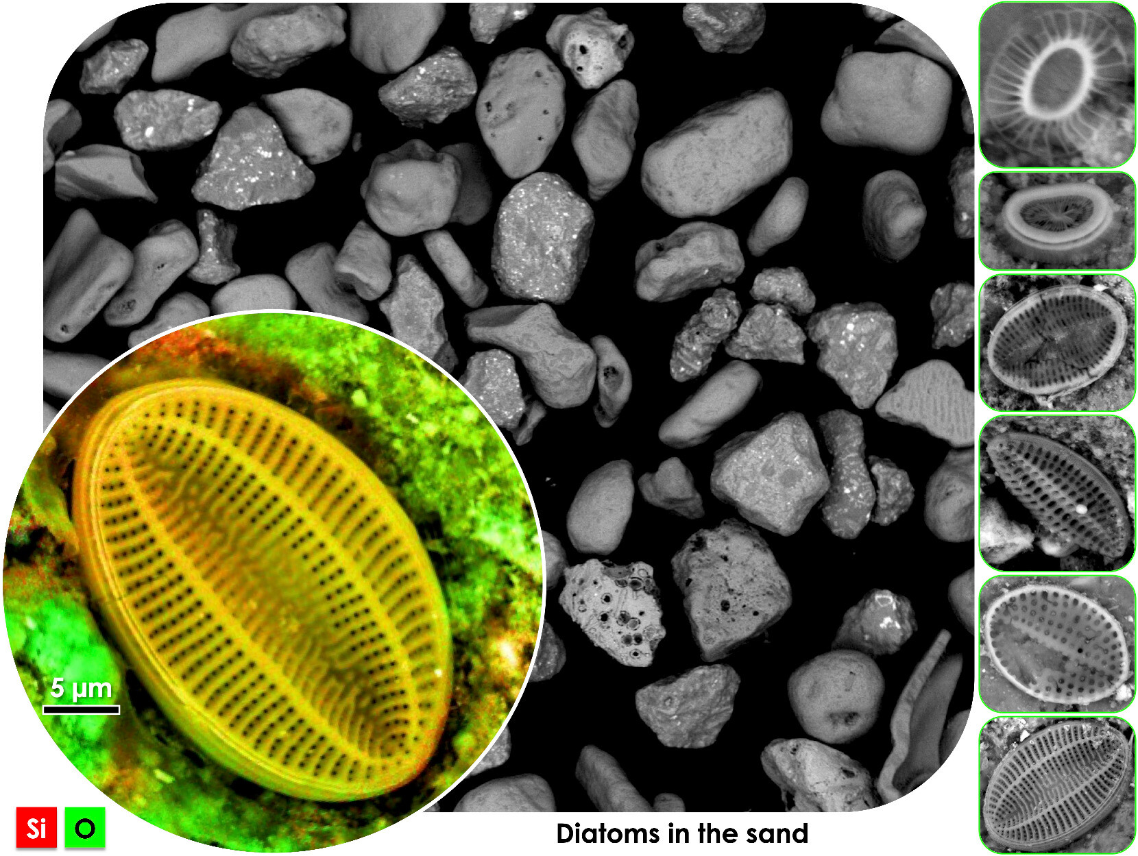
Diatoms on #sand grains visualized using scanning electron microscopy! #Diatoms are a type of microalgae that have intricate and delicate silica shells. Scanning electron microscopy provides incredible detail, allowing us to appreciate the intricacies of #diatom morphology. #ScanningElectronMicroscopy #microscopy #naturephotography #scienceart #microscopicworld #microscopy #biology #lifesciences #silica #science #sciencemastodon #biomineral #biomaterials
– "Strange but beautiful" (@JimiHendrix)
#sand #diatoms #diatom #scanningelectronmicroscopy #microscopy #naturephotography #ScienceArt #microscopicworld #biology #lifesciences #silica #Science #sciencemastodon #biomineral #biomaterials

IT News · @itnewsbot
2898 followers · 250026 posts · Server schleuss.onlineArchaeologists found evidence of trepanation on medieval woman’s skull - Enlarge / This skull of a 50-year-old-ish medieval woman, circa 6th to ... - https://arstechnica.com/?p=1917619 #scanningelectronmicroscopy #historyofmedicine #osteoarchaeology #gaming&culture #bioarchaeology #paleopathology #archaeology #trepanation #ctscanning #science
#science #ctscanning #trepanation #archaeology #paleopathology #bioarchaeology #gaming #osteoarchaeology #historyofmedicine #scanningelectronmicroscopy

Tech news from Canada · @TechNews
273 followers · 6912 posts · Server mastodon.roitsystems.caArs Technica: Archaeologists found evidence of trepanation on medieval woman’s skull https://arstechnica.com/?p=1917619 #Tech #arstechnica #IT #Technology #scanningelectronmicroscopy #historyofmedicine #osteoarchaeology #Gaming&Culture #bioarchaeology #paleopathology #Archaeology #trepanation #CTscanning #Science
#Tech #arstechnica #it #technology #scanningelectronmicroscopy #historyofmedicine #osteoarchaeology #Gaming #bioarchaeology #paleopathology #archaeology #trepanation #CTscanning #science
IT News · @itnewsbot
2389 followers · 242093 posts · Server schleuss.online“Fake” Roman coins authenticated, bearing likeness of lost Roman emperor - Enlarge / This Sponsian gold coin, circa 260-c.270 CE, was part of a ca... - https://arstechnica.com/?p=1903012 #scanningelectronmicroscopy #gaming&culture #romanemperors #romanhistory #spectroscopy #numismatics #romancoins #science #physics
#physics #science #romancoins #numismatics #spectroscopy #romanhistory #romanemperors #gaming #scanningelectronmicroscopy
Paul Harnik · @paulharnik
110 followers · 13 posts · Server ecoevo.social
Beautiful larval shell of the marine bivalve Semelina nuculoides sampled at -20m on the continental shelf offshore FSU's marine lab in the Florida panhandle, imaged today by Luke Calderaro #MolluskMonday #MolluscMonday #ScanningElectronMicroscopy #TinyWorlds
#molluskmonday #MolluscMonday #scanningelectronmicroscopy #tinyworlds
Paul Harnik · @paulharnik
107 followers · 10 posts · Server ecoevo.social
Marine bivalve larval shells from the Florida Panhandle #TinyWorlds #ScanningElectronMicroscopy #Mollusca #Bivalvia
#tinyworlds #scanningelectronmicroscopy #mollusca #bivalvia
Paul Harnik · @paulharnik
95 followers · 6 posts · Server ecoevo.social
Bivalve larval shells from the continental shelf (-20 meters) offshore Alabama, imaged this evening in my lab. Images, clockwise from the upper left, are (1) Parvalucina crenella, (2) Nucula proxima, and (3) Anadara (most likely A. transversa). #TinyWorlds #ScanningElectronMicroscopy
#tinyworlds #scanningelectronmicroscopy